
|Articles|March 12, 2007
CAD technology cools its heels amid skepticism
Does software that flags malignancies on medical images help, hinder, or make no difference to patient management? That question has dogged radiology for years. Automated detection systems are undoubtedly becoming smarter, strengthening arguments for their use. But no system is perfect, and doubts remain, leading to a widespread policy of wait and see.
Advertisement
Does software that flags malignancies on medical images help, hinder, or make no difference to patient management? That question has dogged radiology for years. Automated detection systems are undoubtedly becoming smarter, strengthening arguments for their use. But no system is perfect, and doubts remain, leading to a widespread policy of wait and see.
Computer-aided detection is a good idea in principle, the argument goes, but let's not rush in yet. The technology may still improve further given time. For those who are tired of waiting, speakers at Saturday refresher course showed exactly what CAD can and cannot do.
"The theme that will be manifest in this session is that CAD methods are here, and at least some of them are ready for clinical use," said Dr. James Thrall, radiology chair at Massachusetts General Hospital. "The ones that are not quite ready for clinical use show substantial promise."
CAD software's long-standing weakness is its tendency to flag apparent abnormalities that are not actually malignancies. Progress is being made in this area as the technology evolves. Despite the improvements, however, many radiologists are still not taking advantage of CAD.
"Great improvements can be seen in the latest versions of CAD software for lung CT, making the tool more efficient," said Dr. Catherine Beigelman-Aubry, a radiologist at Pitié-Salpêtrière Hospital in Paris. "But the workflow for CAD systems is not optimized. The best CAD systems are not always available on clinical workstations used for diagnosis."
Systems requiring that CT data sets be transferred to a separate workstation waste too much time, she said. She thinks that lung CAD software will remain underused unless it can be integrated into a reading workstation or PACS.
A number of different lung imaging problems can be addressed with CAD, including detection of pulmonary embolism and quantification of emphysema. Most systems, however, have been designed to pick up lung nodules. The best systems have a sensitivity of around 85% for solid nodules, whereas the sensitivity of a human observer will be around 60%.
"Without a CAD system, screening for lung nodules is a long and tedious task," Beigelman-Aubry said. "Some CAD systems may also depict nonsolid nodules with a higher sensitivity than human observers. This is important to consider because the prevalence of malignancy of these nodules is around 10 times higher than the solid nodules."
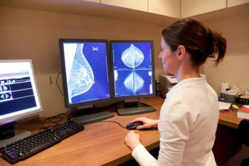
The task of hunting for abnormalities on screening mammograms is fraught with similar problems. Images are complex, and the changes indicative of a cancer can be extremely subtle. Readers faced with a large pile of screening mammograms to report will inevitably become fatigued. Little wonder, then, that there is a reasonably steady miss rate of 20%.
Breast CAD packages are now prevalent in the U.S. Radiologists who previously worked alone can now double-check their findings and reimbursed for doing so. Uptake in Europe, however, has been considerably slower. This is largely due to the long-standing tradition of double reading for breast screening in Europe, said Dr. Rosalind Given-Wilson, a radiologist at St. George's Hospital in London.
As healthcare costs increase, breast screening providers in Europe will undoubtedly look at replacing their second human pair of eyes with a software package. Studies examining the benefits of breast CAD have produced contradictory findings, however. The switch may not even be cost-effective if the number of women called for follow-up rises, but the rate of cancer detection stays reasonably constant.
One area where breast CAD could be used in Europe with relatively little controversy is training. The "show me one like it feature," which brings up similar looking abnormalities and their correct diagnoses, could be a useful tool for trainees, Given-Wilson said. What happens tomorrow will depend on how the technology evolves to reduce false positives and minimize unwarranted recalls.
"At the moment, if given a choice, I would rely on a second reader for screening mammograms. But that's not to say that in a few years time I won't be relying on CAD," she said. "The decision-making interface is where things need to improve. It's not that breast CAD systems are bad, or that radiologists are bad, it is just how they work together."
Newsletter
Stay at the forefront of radiology with the Diagnostic Imaging newsletter, delivering the latest news, clinical insights, and imaging advancements for today’s radiologists.
Advertisement
Related Content
Advertisement


Latest CME


Advertisement
Advertisement
Trending on Diagnostic Imaging
1
Leading Breast Radiologists Discuss the Recent Lancet Study on AI and Interval Breast Cancer
2
Is AI Better Than Neuroradiologists at Evaluating Aneurysm Growth on CTA and MRA Scans?
3
FDA Clears 3T MRI Device for Neonates and Infants
4
FDA Clears AI-Powered Triage Platform for Digital Breast Tomosynthesis
5