
|Articles|November 30, 2004
Contrast ultrasound zeroes in on focal liver lesions
Author(s)H.A. Abella
Sonographic examination with microbubble contrast agents can reliably characterize focal liver lesions, confirm or exclude hepatocellular carcinomas, and remove the need for more invasive, costly imaging, according to studies presented Monday.
Advertisement
Sonographic examination with microbubble contrast agents can reliably characterize focal liver lesions, confirm or exclude hepatocellular carcinomas, and remove the need for more invasive, costly imaging, according to studies presented Monday.
Canadian researchers used contrast-enhanced ultrasound to evaluate 80 patients who presented during a nine-month period with 106 hepatic nodules that had a high likelihood of becoming malignant. The researchers characterized lesions during dynamic and portal vein phase scanning based on the lesions' enhancement patterns. They found that contrast sonography could characterize lesions early and obviate further scanning with CT or MR.
Contrast ultrasound confirmed 46 lesions as HCC, 31 as dysplastic/regenerative nodules, 16 as hemangiomas, eight as pseudomasses, and five as indeterminate nodules. Twenty-one out of 37 nodules detected at initial scanning were benign and were subjected to close surveillance with ultrasound only. None of these lesions evolved into HCC after the nine-month follow-up. Fifteen nodules were diagnosed as HCCs and one as a pseudomass.
Of 69 lesions with an indeterminate diagnosis at initial CT/MR scanning, contrast ultrasound identified 30 HCCs, 19 dysplastic/regenerative nodules, seven hemangiomas, and eight pseudomasses.
In a similar study, Italian researchers enrolled 191 consecutive patients with chronic diffuse liver disease due to chronic hepatitis B or C and cirrhosis. Using microbubble contrast sonography, the researchers accurately differentiated HCCs from other focal liver lesions.
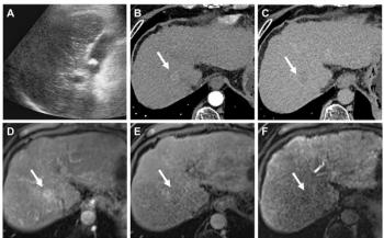
As in the previous study, the investigators used the lesions' enhancement patterns at different phases to calculate positive predictive values. Two independent observers reviewed the studies. HCCs showed 40 diffuse homogeneous, 56 heterogeneous, 10 dotted, and five absent enhancement patterns at arterial phase. HCC also appeared as 71 hypoechoic lesions and 40 isoechoic lesions at late phase.
The investigators obtained the highest positive predictive value for HCCs by combining diffuse heterogeneous enhancement at arterial phase with hypoechoic appearance at late phase, with a PPV of 94% and overall accuracy in HCC characterization of 92%.
Newsletter
Stay at the forefront of radiology with the Diagnostic Imaging newsletter, delivering the latest news, clinical insights, and imaging advancements for today’s radiologists.
Advertisement
Related Content
Advertisement
Latest CME


Advertisement
Advertisement
Trending on Diagnostic Imaging
1
Leading Breast Radiologists Discuss the Recent Lancet Study on AI and Interval Breast Cancer
2
Is AI Better Than Neuroradiologists at Evaluating Aneurysm Growth on CTA and MRA Scans?
3
FDA Clears AI-Powered Triage Platform for Digital Breast Tomosynthesis
4
FDA Clears 3T MRI Device for Neonates and Infants
5