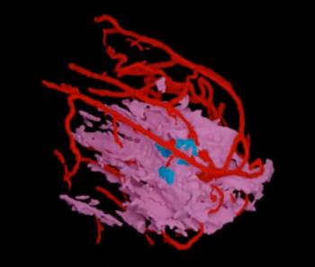
Reducing false positives in evaluation of muscle invasion in bladder cancer cases that can occur with conventional imaging, spectral information with photon-counting CT may offer significantly improved accuracy and specificity in local staging.

Reducing false positives in evaluation of muscle invasion in bladder cancer cases that can occur with conventional imaging, spectral information with photon-counting CT may offer significantly improved accuracy and specificity in local staging.

In the latest episode of his “Interventional Radiology in Focus” series, Mina Makary, MD, discusses the appeal of the specialty, key advances in the field in recent years, and the need for increased awareness of what interventional radiology can offer to patients.

The AI-powered software NeuroQuant PET reportedly offers automated quantification and structured reporting and facilitates evaluation of brain scans with multiple amyloid PET radiotracers.

Preliminary research with the PET radiopharmaceutical 68Ga-NYM096 has demonstrated a 95.7 percent accuracy rate in patients with primary clear cell renal cell carcinoma (ccRCC) and a significantly higher mean SUVmax in the most active lesions in comparison to 18F-FDG.

For patients who had successful reperfusion after acute ischemic stroke, new research demonstrated an 80 percent AUC for predicting infarct with the combination of apparent diffusion coefficient (ADC) mapping and relative oxygen ejection fraction (rOEF).

In a recent Practice Academy webinar session, Nina Kottler, MD, MS, FSIIM, FAIM, discussed challenges with narrow AI applications, the continued emergence of foundation models and a potential trajectory with AI that may facilitate a combination of opportunistic screening and prognostic risk assessment.

In a recent Practice Academy webinar session, Seena Dehkharghani, MD, FAHA, and Raza Mushtaq, MD, discussed the challenge of burgeoning worklist volume amid the ongoing radiologist shortage and the emergence of AI advances with enterprise imaging.

The cmAngio 2.0 AI-enabled software reportedly offers new capabilities for quantitative measurement of breast arterial calcification (BAC) as well as age-based contextualization based on mammography assessment.

New research revealed that risk-stratified MRI surveillance in patients with cirrhosis led to over a 10 percent increase in early detection and over a 44 percent decrease in surveillance exams in comparison to reported results with semiannual ultrasound monitoring.

While maintenance of certification requires participation in peer review, a collegial ethos enables us to provide assessment of another radiologist’s work without crossing the line into odious expert witness cash grabs.

Catch up on the latest contrast-enhanced mammography research, CT insights, MRI studies and FDA approvals in PET imaging with our Weekly Scan.

Recent positron emission tomography (PET) studies with florquinitau F 18 injection (Tauklarify) demonstrated inter-reader agreement ranging between 86 to 92 percent in evaluating patients for Alzheimer’s disease.

The high-resolution temporal and spatial imaging with photon-counting CT bolster our understanding of the connections between anatomy and function in structural heart imaging.

Use of the CARE Multi-Triage CT Body software will be eligible for a New Technology Add-on Payment (NTAP) from CMS starting on October 1, 2026 for inpatients in Medicare fee for service plans.

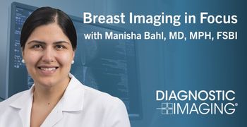
In the latest episode of her “Breast Imaging in Focus” series, Manisha Bahl, MD, MPH, FSBI, offers her perspective on a new study that examines the effectiveness of contrast-enhanced mammography in initial and subsequent rounds of breast cancer screening.

In a recent episode of our “Molecular Imaging in Focus” series, Martin Pomper, MD, PhD, discussed the origin of PSMA PET, shared his perspective on non-PSMA targets in prostate cancer and offered his thoughts on the potential promise of emerging theranostics.

The VueJect ultrasound contrast delivery system, which is geared toward cardiac ultrasound imaging, also gained Health Canada authorization.

Catch up on the latest blogs from Filippo Cademartiri, MD, PhD, who discusses topics ranging from CCTA scan acquisition and photon-counting CT to artificial intelligence and assessment of current plaque as a biomarker for evaluating MACE risk.

In comparison to standard readout-segmented echo-planar imaging, the combination of deep learning reconstruction and multi-shot diffusion-weighted imaging (DWI) offered significantly higher SNR, CNR and lesion contrast with a greater than a two-minute reduction in scan time.

Getting caught up in the numbers of cases we’re reviewing may increase stress and inhibit our productivity.

Catch up on the top radiology content of the past week.

Noting the rising imaging volume and dearth of MRI scanner availability, the authors of a new literature review explore the utility and impact of workflow redesign, staff training, scheduling improvements and AI advances for enhancing MRI workflows.

Smaller voxel size may offer insights into biology we could never access with conventional resolution.

Catch up on the latest episodes of our Breast Imaging in Focus series with Manisha Bahl, MD, MPH, FSBI, discussing research on a variety of topics including breast arterial calcification on mammograms, automated breast ultrasound, AI and GLP-1 receptor agonists.

In a recently published review of the BI-RADS v2025 manual, leading breast radiologists offered pertinent insights on key changes for reporting of mammogram, breast ultrasound and breast MRI interpretation and assessment.

In a recent interview, Kay Jann, PhD, discussed challenges with urinary continence in stroke survivors and a forthcoming NIH-funded study examining the use of brain MRI to help identify those who may benefit from transcutaneous spinal cord stimulation (TSCS).

In comparison to 2023 statistics, the authors of a new study noted an approximately 49 percent increase in radiology turnaround time for ultrasound interpretation and over a 35 percent increase in turnaround time for X-ray assessments.

Extracellular volume fraction (ECV) assessment with photon-counting CT offered higher sensitivity and comparable specificity and AUC to magnetic resonance elastography (MRE)-derived liver stiffness measurement (LSM) for evaluating patients with stage 2 or higher fibrosis, according to a recent study.

New computed tomography (CT) research in July 2026 ranged from the ongoing impact of AI to significant declines in CT radiation dosing over the past decade to a growing recognition that coronary artery calcium (CAC) scoring alone may not be sufficient to convey a true picture of cardiovascular risk stratification.

The AI tools for the newly launched Invenia ABUS Prime ultrasound platform reportedly facilitate a 40 percent increase in scan speed and up to a 93 percent sensitivity rate for lesion detection in women with dense breasts.