Can low-field magnetic resonance imaging (MRI) provide a viable diagnostic alternative to conventional MRI for spinal pathologies?
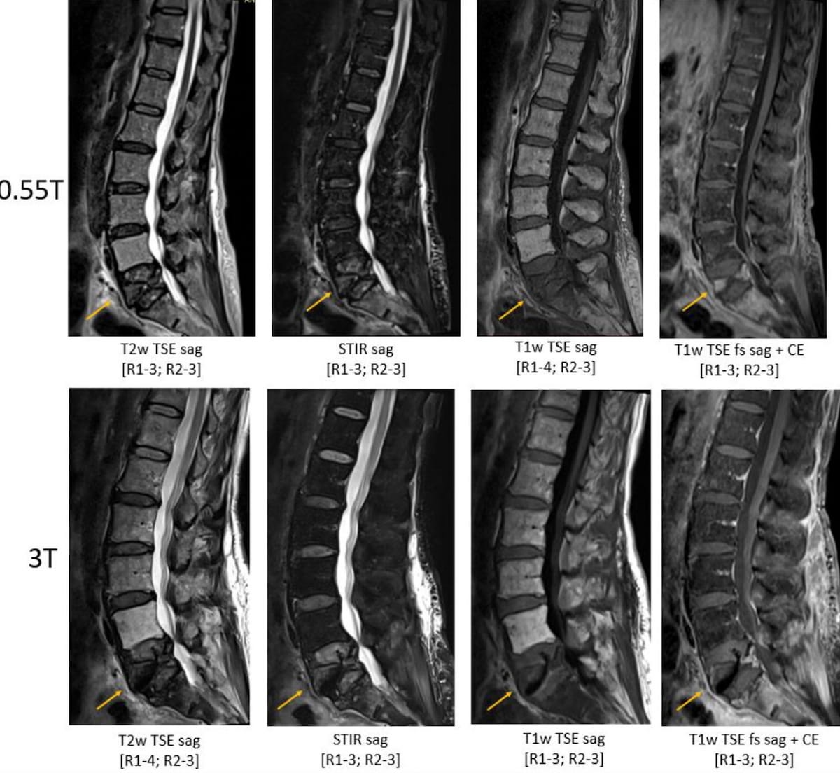
For the retrospective study, recently published in the European Journal of Radiology, researchers reviewed the use of low-field MRI (0.55T) and conventional MRI (1.5T or 3T) in 665 lumbar spine images obtained from 70 studies for 35 patients (59.5 years). The imaging protocol for low-field MRI was comprised of nine sequences including sagittal T2w turbo spin echo (TSE) with spectral fat suppression (fs), sagittal short Tau inversion recovery (STIR), axial (disc spaces) T2w TSE and sagittal T1w TSE fs+CE, according to the study authors.
Using a Likert Scale to assess image quality, the researchers noted two reviewing neuroradiology fellows found that 0.55T MRI provided acceptable diagnostic quality across all reviewed sequences. The study authors noted that angled axial T2w TSE sequence was the only one deemed by both of the reviewers to have significantly lower overall image quality at 0.55T in comparison to 1.5T or 3T MRI. For low-field MRI assessments, the researchers also noted an 86 percent inter-reader agreement on diagnosis.
“These results suggest that clinical spine imaging on commercially available 0.55T systems is feasible and sufficient for evaluating many routine spinal pathologies. Imaging on 0.55T systems offers the potential for numerous benefits, including the potential for reduced cost and easier siting, and may be advantageous in low-resource environments or emergent settings,” wrote study co-author Shruti Mishra, M.D., who is affiliated with the Division of Neuroradiology in the Department of Radiology at the University of Michigan Medical School, and colleagues.
With respect to other imaging aspects, one of the reviewing neuroradiology fellows noted that 0.55T imaging of T2w TSE sagittal and axial views had lower quality than conventional MRI for the majority of assessed features and the second reviewer concurred about inferior 0.55T quality with artifacts, overall image quality and imaging of the neural foramina.
Three Key Takeaways
1. Feasibility of low-field MRI for routine spine imaging. The study suggests that clinical spine imaging using commercially available 0.55T MRI systems is feasible and provides acceptable diagnostic quality for evaluating many routine spinal pathologies.
2. Potential cost-efficiency and diagnostic reliability of low-field MRI. The study highlights the substantial 86 percent inter-reader agreement on diagnosis with low-field MRI at 0.55T, underscoring its reliability. Additionally, the potential for reduced costs and easier siting further positions low-field MRI as a cost-efficient alternative with statistically supported diagnostic capabilities for routine spinal pathologies.
3. Optimization may be needed for specific sequences. While overall diagnostic quality was acceptable, the study identifies a need for additional optimization, particularly for T2-weighted turbo spin echo (TSE) sequences (both sagittal and axial) at 0.55T. Addressing these optimization challenges could enhance the quality of certain imaging aspects for more accurate diagnostic assessment.
“These results suggest that T2w TSE sequences (both sagittal and axial) might benefit from additional optimization at 0.55T,” added Mishra and colleagues.
The study authors also pointed out a 21 percent difference in inter-reader agreement on spinal cord signal assessments (41 percent for 0.55T vs. 62 percent for 1.5T or 3T).
“Spinal cord signal changes are assessed on T2w sequences. Therefore, the consistent lower ratings given by both readers to the T2w TSE sequences at 0.55T may explain this discrepancy,” noted Mishra and colleagues.
(Editor’s note: For related content, see “MRI Study Shows Moderate to Severe Opacities Six Months After COVID-19 Pneumonia for One-Third of Exams,” “Low-field MRI Matches Standard-Of-Care Imaging for Hydrocephalus” and “0.55 MRI Effective in Lung Imaging.”)
Beyond the inherent limitations of a retrospective study, the researchers acknowledged variability with the lumbar spine imaging protocol and that contrast agents were not utilized in several patients who had higher-field MRI scans. The study authors also noted they did not assess the impact of advanced post-processing of low-field MRI images.