


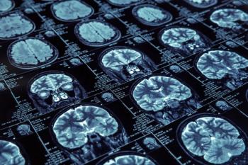
Review top radiology content from the week.

In a recent video interview, Arun Krishnaraj, MD, MPH and David Larson, MD, MBA, discussed the continued use of physical media to transport medical images between different health-care facilities, resulting inefficiencies and delays with patient care, and the initiative to create a linked multi-hub model to end this dated practice once and for all.
Review top radiology content from the week.

What is your diagnosis?
In a recent video interview, Kevin J. Abrams, MD, discussed highlights from the recent 25th Annual Symposium of the American Society of Spine Radiology (ASSR), including lectures on imaging for spontaneous intracranial hypotension, the emergence of dynamic susceptibility contrast for MR perfusion and insights on the use of dual-energy computed tomography (CT) for diagnosing vertebral fractures.

In addition to facilitating strong interreader agreement among radiologists of various experience levels, use of the proposed Neuropathy Score Reporting and Data System (NS-RADS) classification for peripheral neuropathy reportedly improved accuracy in differentiating between milder and more severe categories for muscle abnormalities and nerve lesions.
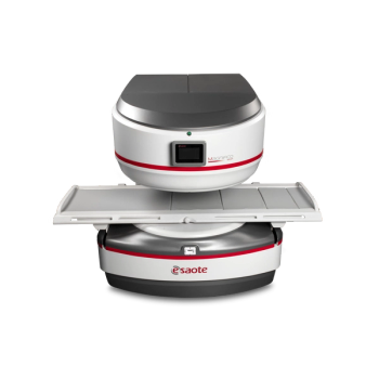
The new whole-body magnetic resonance imaging (MRI) platform reportedly offers enhanced image quality and increased patient comfort.
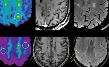
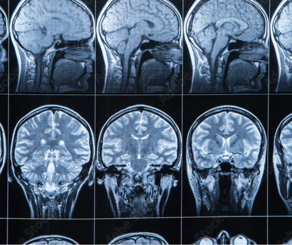
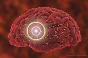
Researchers found the use of magnetic resonance spectroscopic imaging revealed pathologic evidence of multiple sclerosis that is not evident on T1 or T2-weighted MRI.

Expanded MRI labeling for Abbott’s Proclaim™ XR Spinal Cord Stimulation System may facilitate increased efficiency and quality of MRI scans.
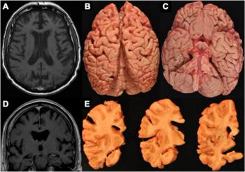
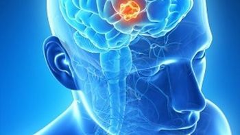
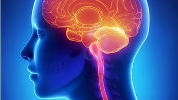
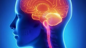
Emerging research suggests that frontotemporal atrophy on magnetic resonance imaging may be a key finding for chronic traumatic encephalopathy (CTE).
New research emphasizes roles of anterior collaterals and ischemic core growth rate in assessing patients with large vessel occlusion.
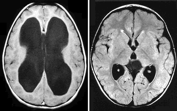
Early study results suggest that low-field MRI may offer a cost-effective, radiation-free alternative to monitor ventricular volume changes in patients with hydrocephalus.
Study findings have the potential to widen the indication for treating patients in the extended window using simpler and more widespread non-contrast CT.
The post-processing software platform can reduce brain imaging acquisition time by 40%.

The algorithm is the first step towards developing an artificial intelligence-augmented radiology workflow that can support image interpretation to improve diagnosis and prognosis.
Combination can also help patients avoid unnecessary invasive procedures.

What is your diagnosis on this MRI?
Use of head and neck CTA has increased significantly among commercially insured and Medicare Advantage patients.

Announcement opens the 10th annual Brain Tumor Segmentation challenge.

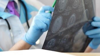
Take a quick look at Diagnostic Imaging's Top 5 stories of the week.
Scans measure blood volume in the brain, assessing oxygen levels and helping to track progress of gene-editing therapies.
An interview with two experts to discuss newly released guidance for using standardized MRI protocols for patients with this condition.
Take a quick look at Diagnostic Imaging's Top 5 stories of the week.
A deep learning algorithm used with brain MRI could help providers identify patients in the early stages of cognitive decline and Alzheimer’s.

Drs. Charles Liu and Jonathan Russin discuss the benefits and potential of fPACT, the alternative to functional MRI.

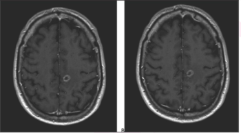
A 30-year-old female presents with papilledema. What is the imaging finding?
Findings could lead to changes in treatment decisions.