
News|Articles|October 3, 2005
64-slice coronary CTA reveals more than stenosis
Contrast-enhanced 64-slice CT can accurately detect and characterize atherosclerotic coronary lesions, according to a study published in the July issue of Journal of the American College of Cardiology. The data signal coronary imaging’s shift in focus from stenosis to coronary plaque.
Advertisement
Contrast-enhanced 64-slice CT can accurately detect and characterize atherosclerotic coronary lesions, according to a study published in the July issue of Journal of the American College of Cardiology. The data signal coronary imaging's shift in focus from stenosis to coronary plaque.
Spatial and temporal resolution drew the boundaries of 16-slice CT's contribution to coronary imaging. Most such CT studies focused on high-grade stenoses in coronary sections larger than 2 mm and in patients with relatively normal or slow heartbeats.
Researchers also failed to integrate catheter angiography's and intravascular ultrasound's capabilities into a noninvasive assessment of ischemic disease using CT. Though 64-slice CT does not overcome all of these issues, its superior resolution provides a thorough analysis of atherosclerotic disease beyond stenosis.
Dr. Alexander Leber and a team of cardiologists and radiologists from the University of Munich in Germany enrolled 59 consecutive patients scheduled for conventional coronary angiography for chest pain. Forty-nine had no previous history of coronary artery disease, while 10 had undergone angioplasty in the past.
The investigators compared 64-slice CT with catheter angiography and intravascular ultrasound to identify and quantify coronary plaque, another departure from previous trials comparing CT exclusively against catheter angiography. They found that 64-slice CT could identify both obstructive and nonobstructive lesions with high accuracy in all three coronaries.
The 64-slice CT scanner provided images of diagnostic quality for the entire coronary tree in 55 of 59 patients. The modality's sensitivity for detection of stenosis of less than 50%, greater than 50%, and greater than 75% was 79%, 73%, and 80%, respectively. Overall specificity was 97%.
CT and intravascular ultrasound identified 46 and 55 coronary plaques, with a sensitivity of 84% and 100%, respectively. The overall correlation between CT, conventional angio, and intravascular ultrasound for the assessment of stenosis degree, mean plaque areas, and percentage of vessel obstruction was statistically significant.
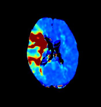
In addition to detection of stenoses, 64-slice CT enables noninvasive analysis of the vessel wall, something that neither catheter angiography nor intravascular ultrasound can do. MSCT is more powerful for analyzing the full spectrum of atherosclerotic disease, whether it causes manifest stenosis or not, said Dr. U. Joseph Schoepf, director of CT research and development at the Medical University of South Carolina.
Symptomatic patients or those with equivocal stress or nuclear examination results undergo 64-slice CT scanning at Schoepf's institution. The implications for patients, medical centers, and the healthcare system are widespread. The high negative predictive value of coronary CTA obviates the need for more invasive, pricey tests. And valuable suite time can be utilized more cost-effectively with patients who truly need intervention, such as angioplasty and stent placement, Schoepf said.
The Munich tudy has limitations. Technical constraints limited the quantification of luminal obstruction, and the sensitivity/specificity values in the study might show a selection bias. Most patients scheduled for catheterization at the German institution usually show up the same day as the procedure, leaving little time for CT.
"It needs to be seen whether future generations of MSCT scanners improve stenosis grading by providing a more comprehensive insight as to the location and nature of atheroslerotic disease," Schoepf said.
For more information from the Diagnostic Imaging archives:
New 64-slice devices drive radiology to new heights
64-slice scanners build case for coronary CTA
Specialists discover common ground in cardiovascular CT
Clinicians weigh 64-slice CT's revolutionary potential
Cardiac imaging: Radiologists move to protect MR and CT turf
Advertisement
Related Content
Advertisement


Advertisement
Advertisement
Trending on Diagnostic Imaging
1
SNMMI: What Early Research Reveals About the Alpha-Emitting Radioconjugate ATNM-400 for Prostate Cancer
2
Mammography Study Shows Impact of AI-Powered Slab Reconstruction with DBT
3
SNMMI: Does Vaccination Enhance Pluvicto Efficacy in mCRPC?
4
SNMMI: Over 80 Percent of Patients with Osseous Oligometastatic PCa are Upstaged with PSMA PET/CT
5