
AI Coverage
about 22 hours ago
Radiology Roundup of FDA News: October 18 — October 24Latest News
Oct 25, 2026 | News
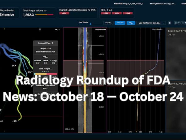
Radiology Roundup of FDA News: October 18 — October 24
Diagnostic Imaging's Weekly Scan: October 18 — October 24
Oct 24, 2026
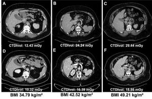
Study Shows PCCT Offers Higher Image Quality and Reduced Radiation Dosing than EID-CT in Obese Patients
Oct 23, 2026
FDA Clears Machine Learning Module for Emerging Musculoskeletal Ultrasound Platform
Oct 23, 2026
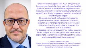
Could Photon-Counting CT Have an Impact as a Platform for Molecular Imaging?
Oct 22, 2026


Advertisement
Shorts
0:49
A Simplified Dosimetry Approach for Treating GEP-NETs: What New Research Reveals
3 days ago
by
Amir Iravani, MD, FRACP

0:34
Can AI Improve Clarity for PI-RADS 3 Assessments on Prostate MRI?
4 days ago
by
Francesco Giganti, MD, PhD

0:47
Theranostics: 'The Biggest Advancement in Our Generation When It Comes to Oncology'
5 days ago
by
Phillip Koo, MD
0:46
Mammography-Based AI Models: Key Takeaways from New Research
7 days ago
by
Manisha Bahl, MD
0:51
How Urinary Excretion Can Facilitate Striking Differences Between PSMA PET Agents
8 days ago
by
Jack Andrews, MD
0:25
Why Prostatic Artery Embolization Should be a First-Line Treatment for Benign Prostatic Hyperplasia
11 days ago
by
Mina S. Makary, MD
0:34
Could Autonomous AI Lead to 'Dramatic Shifts in Compensation' for Breast Radiologists?
13 days ago
by
Wendie Berg, MD, PhD
0:41
Could Future Research Lead to Personalized Treatment with 177Lu-Edotreotide for GEP-NETs?
13 days ago
by
Amir Iravani, MD, FRACP
0:30
Saving AI Output from Mammography Exams Without Causing Unnecessary Patient Worry
17 days ago
by
Manisha Bahl, MD
0:54
Emerging Insights on Embolic Agents for Prostatic Artery Embolization
18 days ago
by
Mina S. Makary, MD
Podcasts
Videos




All News
Advertisement
Advertisement
The future of AI in radiology lies in its potential capability to forge connections and enhance efficiencies for enterprise-wide workflows.

In a new podcast episode, Joseph Cavallo, MD, MBA, offered his perspective on AI integration, potential efficiency gains, navigating post-deployment monitoring and how to incorporate AI literacy into radiology resident training.
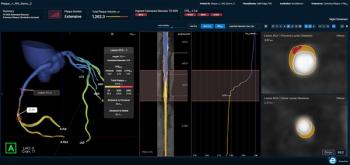
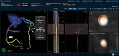
The FFR-IQ software recently demonstrated a 92.2 percent sensitivity for non-invasive estimation of fractional flow reserve (FFRC) based on coronary CT angiography (CCTA) scans.
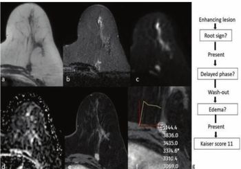
A Kaiser score > 5 with breast MRI demonstrated a 97 percent sensitivity and 87.9 percent specificity for predicting malignancy in patients with BI-RADS 0 or 4 presentations on mammography and breast ultrasound, according to a new study.
In a recent interview, Francesco Giganti, MD, PhD, discussed findings from a recent multicenter study demonstrating the ability of an AI software to provide significant clarity to initial PI-RADS 3 assessments on MRI and facilitate a nearly 19 percent reduction in unnecessary prostate biopsies.
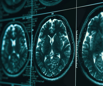
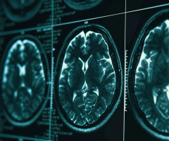
The NEXT-Cue AI software, which incorporates analysis of brain MRI scans, reportedly facilitates prognostic assessments of potential progression from mild cognitive impairment (MCI) to Alzheimer’s disease within five years.
In a new podcast episode, Amir Iravani, MD, FRACP, discusses emerging dosimetry data from the COMPETE trial for 177Lu-edotreotide and how it may help facilitate more personalized treatment for patients with gastroenteropancreatic neuroendocrine tumors (GEP-NETs).
In a recent interview, Phillip Koo, MD, discussed emerging trends with AI, theranostics and empowered patients, and how these developments may lead to challenges and potential opportunities for radiologists and nuclear medicine physicians.
AI has to do more than show potential. It has to deliver measurable results.The Rad AI Outcomes and Market Impact Compendium brings together real-world clinical, operational and financial evidence from health systems, academic medical centers and radiology practices using Rad AI Reporting, Impressions and Continuity.Explore results including 60+ minutes saved per radiologist shift, a 47% reduction in report error rates, 70%+ patient follow-up adherence and >7x ROI — plus, case studies showing how organizations are translating AI into greater capacity, quality and financial performance.See what AI built specifically for radiology can deliver in practice.

Catch up on the top radiology content of the past week.
Advertisement
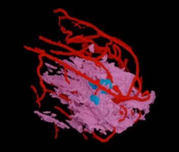
Assessing breast MRI scans, pathology and clinical data, the AI-powered TumorSight Risk software reportedly provides five- and 10-year risk for breast cancer recurrence for women with HR+, HER2-negative stage I-IIIA breast cancer.
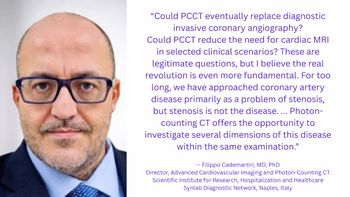
While a new article raises provocative questions about the capability of photon-counting CT (PCCT) to perhaps replace diagnostic ICA, the real revolution with this modality is going beyond stenosis measurement to a renewed focus on the disease of atherosclerosis.
Catch up on the latest imaging research presented at the recent American Society for Radiation Oncology (ASTRO) Annual Meeting.
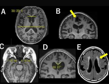
Tightness of the high-convexity sulci (THC) on brain MRI was associated with nearly threefold higher odds of cognitive decline over five to seven years of follow-up, according to a new prospective study involving over 2,100 community-dwelling senior citizens.
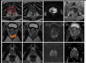
An AI model for prostate MRI offered an 89 percent AUC for prostate cancer detection in external validation testing, according to a newly published international multicenter study.
Advertisement
Advertisement
Trending on Diagnostic Imaging
1
Study Shows PCCT Offers Higher Image Quality and Reduced Radiation Dosing than EID-CT in Obese Patients
2
FDA Clears Machine Learning Module for Emerging Musculoskeletal Ultrasound Platform
3
FDA Clears AI-Enabled FFR-IQ Software for Estimating Fractional Flow Reserve from CCTA
4
Radiology Roundup of FDA News: October 18 — October 24
5