
|Articles|June 16, 2008
Adding 3D software to PACS accelerates workflow
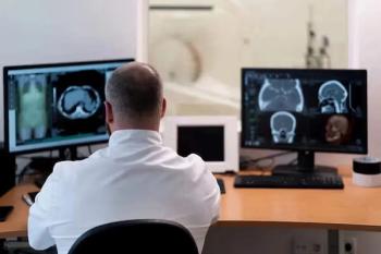
Image-intensive CT and MRI studies can provide radiologists with invaluable diagnostic information-once they have accessed the data-rich studies, that is. Moving large volumetric studies around the hospital network can cause bottlenecks, while time spent loading the images onto a workstation can slow reporting workflows.
Advertisement
Image-intensive CT and MRI studies can provide radiologists with invaluable diagnostic information-once they have accessed the data-rich studies, that is. Moving large volumetric studies around the hospital network can cause bottlenecks, while time spent loading the images onto a workstation can slow reporting workflows.
The Glan Clywd Hospital in Rhyl, U.K., has chosen to tackle these issues by integrating software used to process volumetric studies into the hospital's PACS.
The solution implemented at Glan Clywd is Voxar 3D Enterprise (Barco). Marketed as an advanced visualization solution, it is designed to load 1000+ images in less than 15 seconds through the use of powerful off-the-shelf server-based technology.
"The product has its own separate, short-term storage facility. This lets you pull off the images you want quickly," said Dr. Charles McConnell, lead interventional radiologist at Glan Clywd. "It also has the advantage that when you get a lot of thin slices from multislice CT, you can store them temporarily on the enterprise archive for a few months and then let them go."
Radiologists at the 750-bed district general hospital had already been using an up-to-date version of the Voxar 3D Workstation. This meant that minimal staff training was required for the upgrade. The vendor's 3D Enterprise product has now been installed on 12 workstations in radiologists' offices and departmental reading rooms. Radiologists use the software to reformat thin-slice images and then store these in the PACS ready for hospital-wide access.
"It is very useful for vascular CT, but you can use it for all your routine multislice imaging studies if you want to do multiplanar reconstructions," McConnell said. "We are using it every day, whenever we report CT or MRI examinations."
-By Paula Gould
Advertisement
Related Content


Advertisement
Advertisement
Trending on Diagnostic Imaging
1
FDA Approves Low-Dose MRI Contrast Agent Gadoquatrane
2
Study: PSMA PET/CT Reduces Biopsy Rate by Nearly 50 Percent for Men with Equivocal or Non-Suspicious Prostate mpMRI
3
The Hidden Social Price of Remote Work in Radiology
4
Molecular Imaging in Focus: Heather Jacene, MD, Charts Priorities as the New President of SNMMI
5