AI System Detects Frequently Overlooked Fractures on X-ray
Identifying these easily missed breaks can save patients from future negative complications or loss of function.
A significant number of the most common type of wrist fracture go undetected on X-ray images, setting patients up for potential problems downstream. But, a new artificial intelligence (AI)-based automated system can improve detection rates.
Scaphoid fractures, which typically occur when someone braces from a fall with their hands, are breaks to one of the small bones in the wrist. They account for up to 89 percent of carpal bone fractures and make up approximately 7 percent of all skeletal fractures. They are difficult to see radiologically, though.
“Consequently, scaphoid fractures can be overlooked during initial X-ray examinations,” said study author Nils Hendrix, a doctoral candidate at the Jeroen Bosch Hospital and Jheronimus Academy of Data Science in The Netherlands.
In fact, as many as 50 percent of these breaks can go undetected, leaving up to 12 percent of patients vulnerable to a non-union. If that occurs, individuals can develop avascular necrosis, carpal instability, and osteoarthritis, or they could face surgery for bone re-positioning and fixation or resection of the proximal carpal row. In the most severe circumstances, patient could lose function.

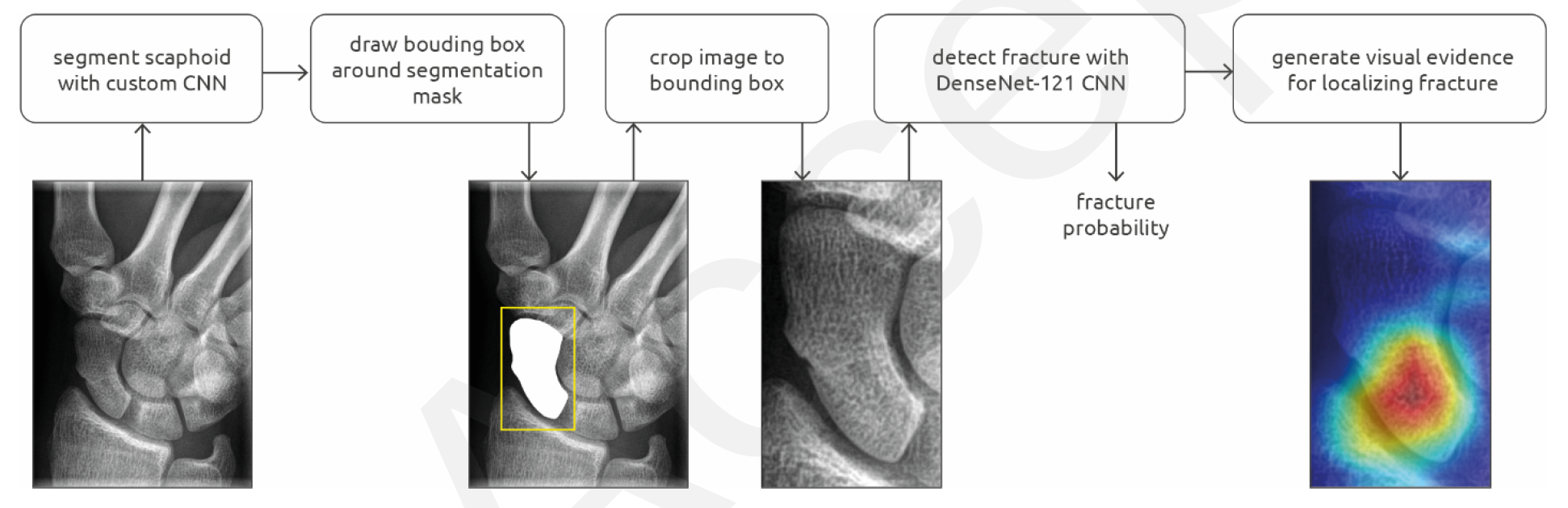
. A class activation map is calculated and visualized as a heatmap for fracture localization.
Credit: RSNA")
Overview of the scaphoid fracture detection pipeline, which consisted of a segmentation and detection convolutional neural network (CNN). A class activation map is calculated and visualized as a heatmap for fracture localization.
Credit: RSNA
But, non-unions can be avoided if fractures are diagnosed within a week and followed by plaster immobilization. X-ray is the imaging modality of choice for detecting these fractures, but the scans can be undermined by variations in wrist position, X-ray technique, or even an overlap of the scaphoid with surrounding wrist bones.
To alleviate this problem, Hendrix’s team developed a deep learning-based system with a convolutional neural network (CNN) that has equivalent diagnostic performance to radiologists in identifying these fractures. They outlined their findings in Radiology: Artificial Intelligence. Their investigation is different from previous work in this area because it is based on a larger data set, as well as algorithm refinements, and it also uses class activation maps that help users pinpoint which image region impacts the network’s predictions.

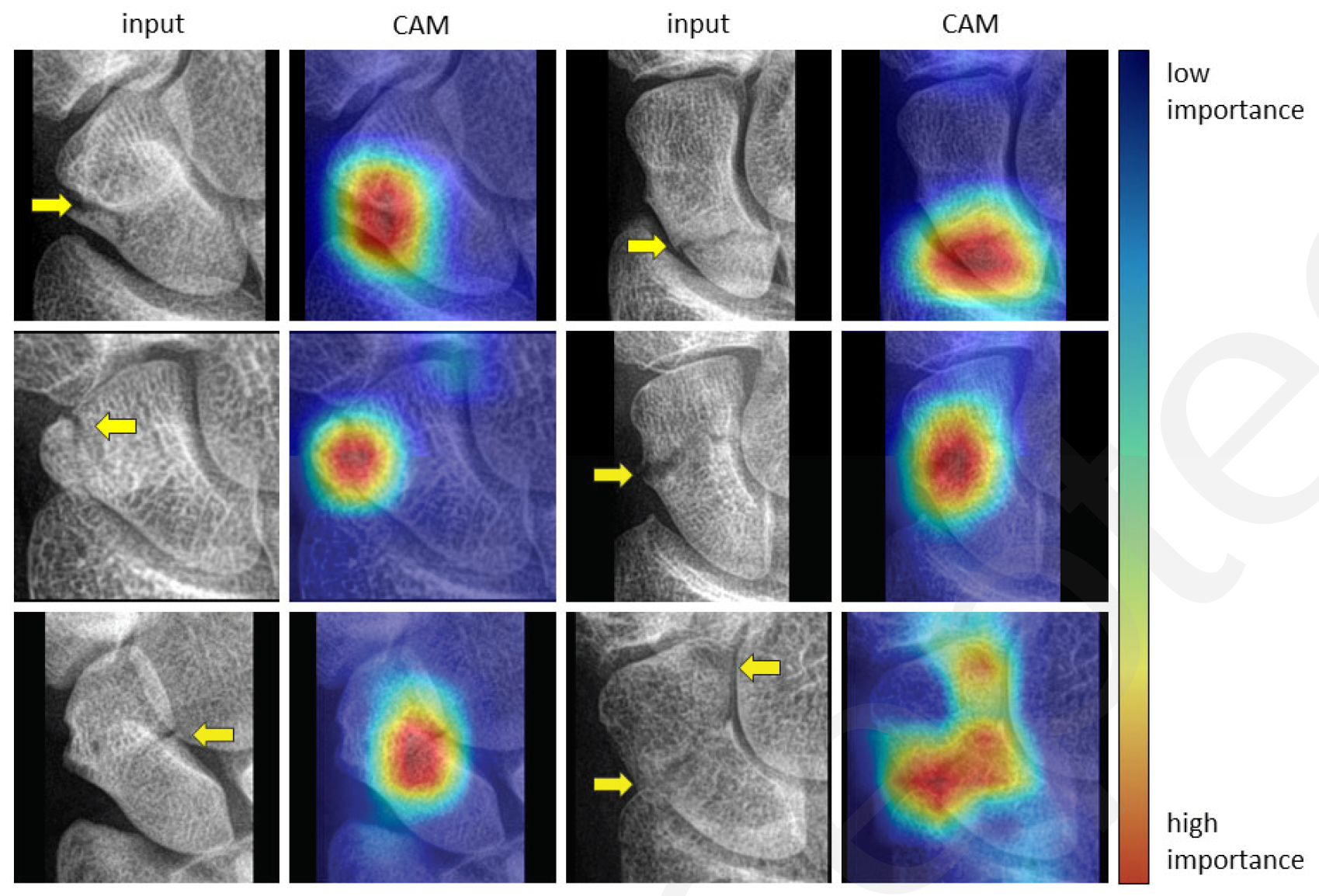
 for localizing fractures. The interpretation of the CMAs follows from the color coding of a heat map, in which pixel regions with a warm color signify a greater influence on the final decision of the network than regions with a cold color. The yellow arrows projected on the input images indicate the fracture lines, and they are only shown for reference.
Credit: RSNA")
Examples of class activation map (CAM) for localizing fractures. The interpretation of the CMAs follows from the color coding of a heat map, in which pixel regions with a warm color signify a greater influence on the final decision of the network than regions with a cold color. The yellow arrows projected on the input images indicate the fracture lines, and they are only shown for reference.
Credit: RSNA
For their study, they used 1,039 conventional hand, wrist, and scaphoid X-rays to develop a scaphoid segmentation CNN and 3,000 X-rays to develop a scaphoid fracture detection CNN. To test the complete fracture detection system, they used 190 X-rays and compared the performance to 11 radiologists.
The radiologists assessed the scans for scaphoid fracture and rated their confidence on a 0-to-1.0 scale with 1.0 indicating absolute certainty.
According to their analysis, the segmentation CNN reached a Dice similarity coefficient of 97.4 percent, and the detection CNN had 78-percent sensitivity, 84-percent specificity, and 83-percent positive predictive value. Their evaluation also showed no difference between the area under the curve of the CNN and the radiologists – 0.87 and 0.83, respectively.
The CNN produced four false-negatives and 14 false positives not made by the average radiologist. It missed one fracture and mis-classified six non-fractures as breaks. The average radiologist made 18 false-negatives and one false-positive not made by the CNN, and with high certainty, the average radiologist missed five fractures.
Based on these outcomes, Hendrix said, the CNN and the class activation maps that overlapped with fracture lines in the scaphoid have great potential for clinical use.
“The system may be able to assist residents, radiologists, or other physicians by acting either as a first of second reader, or as a triage tool that helps prioritize worklists, potentially reducing the risk of missing a fracture,” he said.
Specifically, he said, the tool could reduce the incidence and cost of additional imaging and unnecessary therapy, accelerate diagnosis, and facilitate earlier treatment. In addition, it could prevent delayed therapy and reduce complications that could open the door for a negative clinical outcome.
“The convolutional neural network may also reduce unnecessary wrist immobilization performed out of precaution, in more than half of the patients with clinical suspicion for having a scaphoid fracture,” he added.
Hendrix’s team is now working to expand the scaphoid fracture detection system so it can analyze multiple X-ray views in order to make predictions. The ultimate goal, they said, is to expand the system to other bone structures.
For more coverage based on industry expert insights and research, subscribe to the Diagnostic Imaging e-Newsletter here.


Considering Breast- and Lesion-Level Assessments with Mammography AI: What New Research Reveals
June 27th 2025While there was a decline of AUC for mammography AI software from breast-level assessments to lesion-level evaluation, the authors of a new study, involving 1,200 women, found that AI offered over a seven percent higher AUC for lesion-level interpretation in comparison to unassisted expert readers.


