Alleviating Patient Imaging Fears During COVID-19
Radiology department staff have the ability – and responsibility – to calm patient anxieties about imaging studies and the virus during the pandemic, says one radiology supervisor.
The number of patients nationwide who have undergone some type of diagnostic imaging – chest X-ray or chest CT – during the COVID-19 pandemic currently is not known, but the volume is continuing to rise. For many, that scan could be their first experience in the radiology department, and it can be frightening.
A nervous patient is frequently at jittery patient, and that can mean any images you gather could be of lower quality for diagnosis. Consequently, it can be in the best interest of all parties – patient, technologist, and you and your colleagues – to identify ways to allay the patient’s concerns.
According to Randi Homan, BS, RT, multi-modalities supervisor at Novant Health Huntersville Medical Center in North Carolina, COVID-19 patients are not only scared about viral infection and what the imaging scan might entail, but they can also be nervous about being viewed and treated differently than any of your other patients.
“A lot of patients are expressing to us a fear of being labeled or recognized as a certain dangerous patient population," she siad. "And, they’re nervous about that.”
Not only are patients concerned about potentially being intubated and placed on a ventilator, she said, but they also share concerns about the process of having an imaging scan done. They worry it might hurt or take an inordinate amount of time. In many cases, radiology staff at Novant spend several minutes prior to the study explaining the scans are only a detailed view of the chest and lungs designed to ensure that COVID-19 is not spreading or presenting as some other illness or pathology.
To help put people at ease, Homan said, she and her staff focus on both the educational and emotional aspects augmenting patient comfort. When a patient arrives for a scan, she said, the technologist provides a detailed description of the scan the referring provider has ordered – what will happen, why the doctor requested the study, and the importance of examining the patient’s heart and lungs.
In addition, she stressed, you must endeavor to connect with the patient. Be friendly, and use your body language to help the patient maintain calm.
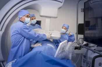
“In our department, we wear a great deal of personal protective equipment, but that prevents the patient from really being able to see us, especially our mouths,” she explained. “So, I make a conscious effort to try to smile with my eyes.”
It can be helpful to explain that you’re wearing personal protective equipment for both your safety and theirs. Homan also recommended maintaining eye contact as much as possible, as well as speaking clearly and offering words of affirmation. If possible, providing a warm blanket prior to the scan can help some patients relax.
Ultimately, she said, the radiology department can – and should – play a role in helping patients navigate through a difficult time.
“You really need to do everything possible to help ensure the patient’s safety and their comfort. Remember to keep an open mind with them,” she said. “Many are being categorized as the ‘COVID-19’ patients, but they have their own unique personalities, family and friends. We need to go above and beyond to ensure their safety, comfort, and confidence.”
Newsletter
Stay at the forefront of radiology with the Diagnostic Imaging newsletter, delivering the latest news, clinical insights, and imaging advancements for today’s radiologists.


