Assessing the Potential of Positron Emission Mammography: An Interview with Vivianne Freitas, MD
In a recent interview, Vivianne Freitas, M.D., discussed new research findings on positron emission mammography (PEM), pertinent benefits of the technology and its potential as a viable alternative in the future to conventional imaging techniques for breast cancer screening.
Recent pilot study results from the use of low-dose positron emission mammography (PEM) revealed a 96 percent detection rate for malignant breast lesions as well as an 87.1 percent sensitivity rate and a 94.7 percent specificity rate. The researchers also found that PEM was effective regardless of breast density and had a lower percentage of false positives than breast MRI, according to the study, which was recently published in Radiology: Imaging Cancer.
In a recent interview, lead study author Vivianne Freitas, M.D., who is affiliated with the Joint Department of Medical Imaging with the University Health Network and the Sinai Health System at the Women’s College Hospital at the University of Toronto in Canada, shared her thoughts about the study findings and the future potential of PEM.
Q: What sparked your interest in the PEM technology?
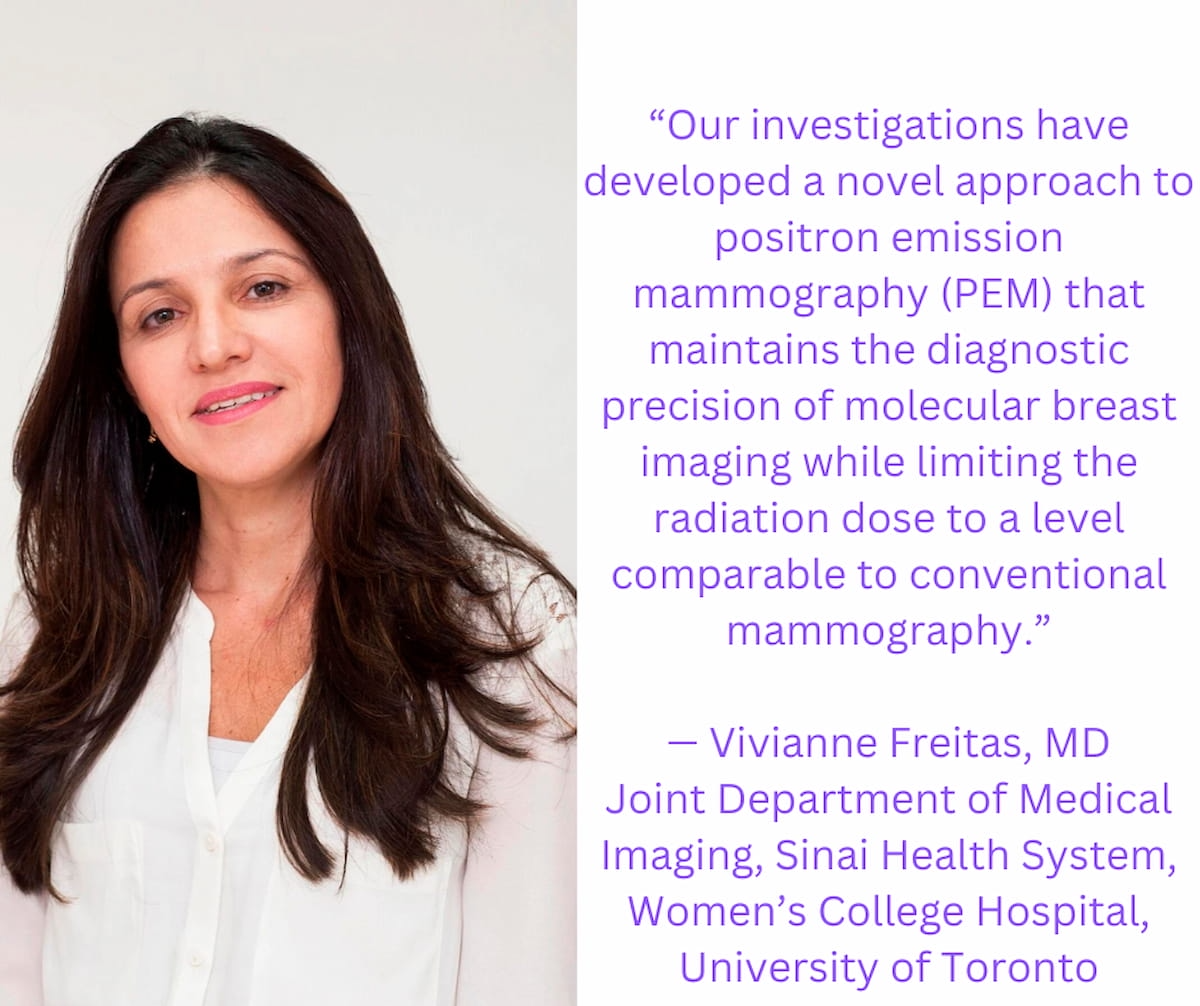
A: My academic goals revolve around advancing novel imaging methods to improve cancer detection at its early stages. PEM technology stands out as a particularly promising avenue in this field. It operates by administering a radioactive tracer preferentially absorbed by cancerous cells over healthy ones, highlighting these cells as distinct "hot spots" in the imaging results. Although molecular breast imaging (MBI) is an effective technique, its significant limitation has been the high level of radiation exposure it necessitates. Nevertheless, our investigations have developed a novel approach to positron emission mammography (PEM) that maintains the diagnostic precision of MBI while limiting the radiation dose to a level comparable to conventional mammography.

Q: What are the potential advantages of PEM compared to mammography and MRI?
A: This innovative imaging modality aims to integrate the high sensitivity of MRI while significantly reducing the likelihood of false positives. One of its key advantages is its low cost compared with MRI, making it a more accessible option for widespread use. Additionally, this technology is designed to deliver a radiation dose comparable to that of traditional mammography. However, unlike mammography, it achieves this without the need for breast compression, which can often be uncomfortable for patients.
The technology also stands out for its efficacy regardless of breast density, a critical factor since dense breast tissue can obscure the results of standard screening methods like mammography. By addressing this challenge, the new imaging solution promises to enhance breast cancer screening's reliability for a wider patient demographic, especially those with dense breasts where early detection poses greater difficulty.
Combining these attributes — improved sensitivity, reduced risk of false positives, economic viability, acceptable radiation exposure without compression, and effectiveness independent of breast density — this novel imaging strategy emerges as a potentially revolutionary breakthrough in early breast cancer detection. It has the potential to reshape breast cancer diagnostics and screening, offering a complement or even an improvement over existing techniques and representing a significant advancement in the management of breast cancer.
Q: What are some of the key benefits of PEM for patients?
A: Our team conducted a pilot study involving 25 female participants, all diagnosed with breast cancer, with a median age of 52. The participants underwent low-dose PEM using the radiotracer fluorine 18-labeled fluorodeoxyglucose (18F-FDG) images. Captured one and four hours after the injection of 18F-FDG, these images were analyzed by two specialized breast radiologists and compared against laboratory results. This method demonstrated high accuracy, identifying 24 out of the 25 invasive breast cancers, translating to a 96 percent detection rate. Notably, its false positive rate was a mere 16 percent, lower than the 62 percent associated with MRI scans.
This advancement could profoundly affect patient care by lowering patient anxiety and decreasing the number of unnecessary biopsies and treatments. It represents a step forward toward a more focused and accurate approach in the medical field.
Q: From your perspective, can PEM be a viable alternative in terms of cost-effectiveness?
A: Despite these promising results, pursuing additional research to delineate its practical applications in real-world settings is crucial. Future studies, including cost-effectiveness studies, must determine its definitive role and effectiveness within the clinical setting.
Considering Breast- and Lesion-Level Assessments with Mammography AI: What New Research Reveals
June 27th 2025While there was a decline of AUC for mammography AI software from breast-level assessments to lesion-level evaluation, the authors of a new study, involving 1,200 women, found that AI offered over a seven percent higher AUC for lesion-level interpretation in comparison to unassisted expert readers.
The Reading Room Podcast: Current Perspectives on the Updated Appropriate Use Criteria for Brain PET
March 18th 2025In a new podcast, Satoshi Minoshima, M.D., Ph.D., and James Williams, Ph.D., share their insights on the recently updated appropriate use criteria for amyloid PET and tau PET in patients with mild cognitive impairment.