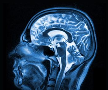
Brain MRI of People with Genetic Autism
MRI has shown structural abnormalities related to cognitive function among people with genetic forms of autism.
MRI has identified structural abnormalities in the brains of people with genetic autism, according to a study published in
Researchers from California, New York, Texas, Pennsylvania, and Massachusetts sought to identify developmental neuroradiologic findings in carriers who have deletion and duplication at 16p11.2, one of the most common genetic causes of autism spectrum disorder (ASD), and to assess how these features are associated with behavioral and cognitive outcomes.
The study included:
• 79 deletion carriers (42 male), ranging in age from 1 to 48 years, mean age 12.3
• 79 duplication carriers (43 male), aged 1 to 63 years, mean age 24.8
• 64 unaffected family members (31 male), aged 1 to 46 years, mean age 11.7 years
• 109 participants (64 male) in a control group, aged 6 to 64 years, mean age 25.5
All subjects underwent structural MR imaging and completed cognitive and behavioral tests. Neuroradiologists reviewed the MR images for development-related abnormalities. “People with deletions tend to have brain overgrowth, developmental delays and a higher risk of obesity,” study author Julia P. Owen, PhD, a brain researcher at the University of Washington in Seattle, said in a release. “Those with duplications are born with smaller brains and tend to have lower body weight and developmental delays.” Owen was at the University of California in San Francisco (UCSF) during the study.
The most prominent features of those who carried the deletion compared with familial noncarriers and population control participants were found to be:
•More prominent dysmorphic and thicker corpora callosa (16%) for carriers
• Greater likelihood of cerebellar tonsillar ectopia (30.7%) for carriers
• Chiari I malformations (9.3%) for carriers
For duplication carriers, the most salient findings compared with familial noncarriers and population control participants were:
• Reciprocally thinner corpora callosa (18.6%) for carriers
• Decreased white matter volume (22.9%) for carriers
• Increased ventricular volume (24.3%) for carriers[[{"type":"media","view_mode":"media_crop","fid":"62557","attributes":{"alt":"","class":"media-image media-image-right","id":"media_crop_9243723493469","media_crop_h":"0","media_crop_image_style":"-1","media_crop_instance":"7944","media_crop_rotate":"0","media_crop_scale_h":"0","media_crop_scale_w":"0","media_crop_w":"0","media_crop_x":"0","media_crop_y":"0","style":"height: 445px; width: 300px; float: right;","title":"Example images for a control participant , a deletion carrier, and a duplication carrier. In the sagittal image of the deletion carrier, the thick corpus callosum, dens and craniocervical abnormality, and cerebellar ectopia are shown. For the duplication carrier, the sagittal image shows the thin corpus callosum and the axial image shows the increased ventricle size and decreased white matter volume. ©RSNA 2017 ","typeof":"foaf:Image"}}]]
Results of the cognitive assessments compared to the imaging findings showed the presence of any imaging feature associated with deletion carriers resulted in worse daily living, communication, and social skills compared with deletion carriers without any radiologic abnormalities. For the duplication carriers, the presence of decreased white matter, callosal volume, and/or increased ventricle size was associated with decreased full-scale and verbal IQ scores compared with duplication carriers without these findings.
The researchers concluded that there were reciprocal neuroanatomic abnormalities among two genetically related cohorts at high risk for autism spectrum disorder, and determined to be associated with cognitive and behavioral impairments.
“Often studies like this focus on high-functioning individuals, but this was an ‘all-comers’ group,” senior author Elliott Sherr, MD, PhD, head of the Brain Development Research Program at UCSF, said in the release. “We didn’t do mathematical algorithms but rather used the trained eyes of neuroradiologists to evaluate the scans of a full range of individuals. When you look at a broad range of people like this, from developmentally normal to more significantly challenged, you’re better able to find these correlations.”
Newsletter
Stay at the forefront of radiology with the Diagnostic Imaging newsletter, delivering the latest news, clinical insights, and imaging advancements for today’s radiologists.


