Brain Pattern Alterations Found Among Children with Autism Spectrum Disorder
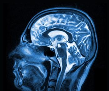
Magnetic resonance imaging shows brain pattern alterations among pre-school children with autism spectrum disorder.
Preschoolers with autism spectrum disorder (ASD) have significantly altered patterns of global and local brain network topography as seen by diffusion-tensor imaging and T2-weighted imaging on a 3-T magnetic resonance
system, according to a study published in the journal Radiology.
Researchers from China performed a small study to investigate this topologic architecture, comparing 21 pre-school children with ASD with 21 children who have typical development (TD). The mean age of all children was 4.5 years. The children with ASD were diagnosed according to the Diagnostic and Statistical Manual of Mental Disorders Global Assessment of Functioning score. All children underwent imaging.
The results showed that compared with the TD group, children with ASD demonstrated shortened characteristic path length and increased global efficiency, and clustering coefficient. Significant increases in nodal efficiency
were mainly found in left pallidum and right caudate nucleus of the basal ganglia network.
“Altered brain connectivity may be a key pathophysiological feature of ASD,” study co-author Lin Ma, MD, from the Department of Radiology at Chinese PLA General Hospital in Beijing, said in a release. “This altered connectivity is visualized in our findings, thus providing a further step in understanding ASD.”
The researchers concluded significantly altered patterns of global and local brain network topography may underlie the abnormal brain development in preschool children with ASD compared with those who have TD. These findings may point toward potential imaging biomarkers for preschool-age patients with ASD.
In the release, Ma explained that in the future, this type of brain imaging might aid in the delivery of ASD therapies for children, like repetitive transcranial magnetic stimulation, and transcranial direct current stimulation. Both are being investigated as possible treatments for ASD.
Newsletter
Stay at the forefront of radiology with the Diagnostic Imaging newsletter, delivering the latest news, clinical insights, and imaging advancements for today’s radiologists.


