In a multicenter study of over 800 women with invasive breast cancer (IBC), an ultrasound-based nomogram combining deep learning and radiomics had a 16 percent higher AUC and over a 30 percent higher sensitivity rate than a clinical model for predicting lymphovascular invasion (LVI).
For the retrospective study, recently published in Academic Radiology, researchers compared a clinical model, B-mode ultrasound (BMUS), and color Doppler flow imaging (CDFI) to the aforementioned deep learning radiomics nomogram (DLRN) for predicting lymphovascular invasion in 832 women with IBC. The DLRN model incorporated tumor margin, tumor size, BMUS score, CDFI score and ultrasound-reported axillary lymph node metastasis (ALNM), all of which were proven to be independent LVI predictors, according to a multivariate analysis within the study.
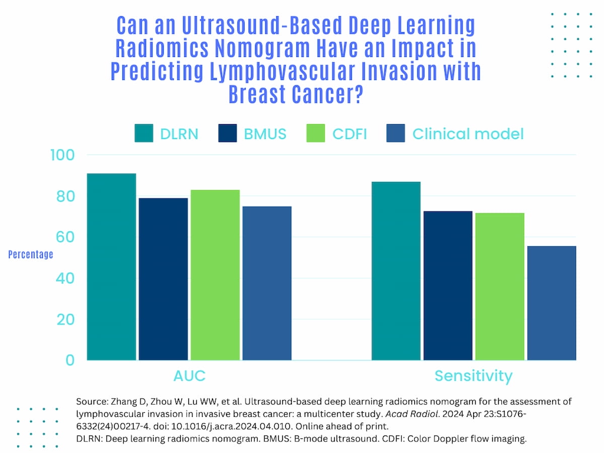
In external validation testing, the researchers found that the DLRN model had a 91 percent area under the receiver operating characteristic curve (AUC) for predicting LVI in comparison to CDFI scoring (83 percent), BMUS scoring (79 percent) and a clinical model (75 percent).
The DLRN model also had a sensitivity rate (86.9 percent) that was over 30 percent higher than that of the clinical model (55.6 percent) and over 14 percent higher than BMUS scoring (72.7 percent) and CDFI scoring (71.7 percent), according to the study authors.
“The user-friendly graphical tool developed in this research has the potential to facilitate non-invasive assessment of LVI status, providing valuable insights into enhancing clinical decision-making processes,” wrote study co-author Chao-Xue Zhang, M.D., who is affiliated with the Department of Ultrasound at the First Affiliated Hospital of Anhui Medical University in Anhui, China, and colleagues.
“To the best of our knowledge, this study was the first attempt to investigate the feasibility of applying a deep learning radiomics approach based on BMUS and CDFI images to assess LVI status in IBC.”
The DLRN model also demonstrated a nearly 20 percent higher negative predictive value (NPV) (85.9 percent) in comparison to the clinical model (66.4 percent) in external validation testing, according to the researchers.
Three Key Takeaways
- Enhanced prediction accuracy. The ultrasound-based nomogram, utilizing deep learning and radiomics, exhibited significantly higher predictive performance for lymphovascular invasion (LVI) in invasive breast cancer (IBC) compared to conventional clinical models. This suggests that incorporating advanced imaging techniques can significantly improve the accuracy of LVI prediction, aiding in more precise clinical decision-making.
- Improved sensitivity and negative predictive value. The deep learning radiomics nomogram (DLRN) not only showed a 16 percent higher area under the receiver operating characteristic curve (AUC) but also demonstrated over 30 percent higher sensitivity and nearly 20 percent higher negative predictive value (NPV) compared to the clinical model. This indicates that DLRN has the potential to identify more cases of LVI accurately and reliably exclude cases without LVI, thereby reducing false negatives.
- Multi-channel information integration. By incorporating multiple ultrasound modalities (B-mode ultrasound and color Doppler flow imaging) and utilizing radiomic feature extraction, the DLRN model surpasses conventional imaging markers. This multi-channel approach helps mitigate challenges associated with tumor heterogeneity, enhancing prediction accuracy. This highlights the importance of integrating diverse imaging data sources for more comprehensive assessment and prediction in breast cancer diagnostics.
Noting that prior radiomics studies for predicting LVI heavily relied upon BMUS scoring, the study authors said the incorporation of BMUS and CDFI, and radiomic feature extraction from those imaging modalities go beyond conventional imaging markers and provide some prognostic information that may be “imperceptible” to visual review.
“The ability to make decisions based on multi-channel information from a tumor mitigates the challenges associated with tumor heterogeneity, thus enhancing prediction accuracy,” emphasized Zhang and colleagues.
(Editor’s note: For related content, see “Whole Breast Ultrasound Screening: Is There Adequate Utilization in Patients at Higher Risk for Breast Cancer?,” “Ultrasound Nomogram May Enhance Prediction of Metastatic Axillary Lymph Nodes in Breast Cancer Patients” and “AI and Breast Ultrasound: Where Things Stand.”)
Beyond the inherent limitations of a retrospective study, the authors acknowledged that extrapolation of the findings to broader populations may be limited with the multicenter cohort entirely comprised of participants from China. The researchers conceded an emphasis on conventional ultrasound modes and noted the data came from various ultrasound units utilized in different facility settings.