
|Articles|April 17, 2013
CAD-enhanced Mammo Gives More Early Cancer False Alarms
Author(s)Diagnostic Imaging Staff
Screening mammograms enhanced by CAD trigger more false alarms than non-CAD screenings and detect more early cancers that may never have needed treatment.
Advertisement
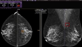
Mammograms enhanced with computer-aided detection (CAD) programs are detecting more noninvasive and early-stage breast cancers, but are also causing more false alarms, according to a study in the
As use of CAD during screening mammography has increased among Medicare beneficiaries, so has the diagnosis of ductal carcinoma in situ (DCIS) and the incidence of diagnostic testing among women who do not have breast cancer.
“DCIS progresses slowly, if at all. Some of these early noninvasive lesions may never have come to clinical attention in women’s lifetimes if CAD were not applied to their mammograms,” lead author Joshua Fenton, MD, MPH, said in a release.
Fenton and his colleagues from the University of California Davis Health System undertook a retrospective cohort study of Medicare enrollees to determine associations between use of CAD with mammography and the incidence of invasive breast cancer, and invasive cancer stage and resultant diagnostic testing.
The researchers assessed 409,459 mammograms from 163,099 women, aged 67 to 89, who underwent screening between 2001 and 2006. The researchers were looking for incidences of DCIS and invasive breast cancer within one year of the mammography, as well as invasive cancer stage and any diagnostic testing performed within 90 days of the screening among the women who did not have breast cancer.
Researchers found that women whose screening included CAD had a 17 percent increase in the diagnosis of DCIS breast lesions and a 6 percent increase in diagnosis of early-stage invasive breast cancer. However, there was also a 19 percent increase in diagnostic mammography and breast ultrasonography after initial screening and a 10 percent increase in breast biopsies.
“CAD is expensive technology that has been nearly universally adopted in the U.S. due to Medicare’s support and the hope that it can help us identify and treat invasive breast cancer early,” Fenton says. “Our study suggests that we still don’t know whether the benefits outweigh the harms for the average woman on Medicare.”
Advertisement
Related Content
Advertisement


Advertisement
Advertisement
Trending on Diagnostic Imaging
1
What Does the Future Hold for Nuclear Medicine?
2
Top Five Radiology Content in June 2026
3
What New MRI Research Reveals About Endometrial Cancer Staging and the 2023 FIGO Staging System
4
FDA Clears New CEM Image Reconstruction and Biopsy Capabilities for Siemens Healthineers Mammography Device
5