
|Articles|April 2, 2006
Cardiology PACS must produce moving images
Tensions between radiology and cardiology in the field of cardiac imaging have persisted for decades. The 1970s and 1980s were rife with turf battles over interventional angiography, echocardiography, and cardiac SPECT. More recently, cardiac MRI and CT have been the focus of intense debate about who is best qualified to perform and interpret these scans.
Advertisement
Tensions between radiology and cardiology in the field of cardiac imaging have persisted for decades. The 1970s and 1980s were rife with turf battles over interventional angiography, echocardiography, and cardiac SPECT. More recently, cardiac MRI and CT have been the focus of intense debate about who is best qualified to perform and interpret these scans.
Passion on both sides has been fueled, at least in part, by concerns about money. But radiologists and cardiologists also often have fundamentally different underlying philosophies about basic issues such as the role of technology and approaches to diagnostic testing. In many respects, these cultural differences and the ways in which they manifest themselves are the same today as they have been for many years.
Yet, whether we fully recognize it or not, we are now entering an era in which an entirely new force is beginning to shape the future of cardiac imaging. At issue is not simply another imaging technique based on a different physical principle. Rather, the issue is the manner by which the image data themselves are stored and accessed by PACS. It seems inevitable that image management will ultimately become part of the larger effort to create a nationwide system for electronic medical records, but questions remain.1-3 Are radiologists and cardiologists on the same page with respect to PACS and the EMR? How will the old cultural differences manifest themselves in the new field of electronic information management? These differences are already beginning to express themselves in cardiac PACS.
It is not uncommon to hear stories of radiology groups that have invested in hardware and software upgrades for new MRI and CT techniques, specifically to allow cardiac imaging, only to be disappointed by the lack of patient referrals from their colleagues in cardiology. One possible explanation is that cardiologists have a hidden agenda that they themselves would like to perform these procedures. In some cases, this may be true, but the real reason for a lack of referrals may be completely different: Cardiologists want to see the pictures. And they want to see them right now.
Consider the case of cardiac MRI and/or cardiac CT. The physical size of the scanner and the intrinsic siting requirements virtually guarantee that the scanner will be located far from everyday cardiology services. At the same time, the fact that these procedures are new also guarantees that referring physicians will have had little experience with them. In many cases, these physicians may never have even seen this type of image in their own patients. This situation will prompt a strong desire to see the images firsthand. Yet, for decades, the only realistic mechanism for seeing the images has been to walk to the scanner, spend favors asking to access someone else's workstation, and then give up in frustration after realizing that the only person who actually knows how to push all of the workstation buttons in the proper order and thus display the images is "unfortunately, not here right now."
What is needed today is essentially the same thing that has been needed for the past 20 years: remote access to cardiac images.4 Equipment manufacturers now enthusiastically assert that their systems can be used to view cardiac images in a practical manner without physically visiting the scanner facility. This type of remote access, particularly if it is made available immediately following the scan, has the potential to directly influence the success and even the shape of cardiac imaging services.
If the cardiologist can see the images right away, he or she may be happy to send patients to radiology. If not, cardiologists almost certainly will not order these tests routinely and might even decide to begin performing the new procedure themselves. Thus, the success or failure of a cardiology PACS may have a direct and profound effect on both the rate of adoption of new imaging technologies and the relative balance of radiology and cardiology in ordering, performing, and interpreting cardiac MR and CT images.
IMAGE MANAGEMENT 101
The heart moves continuously. This deceptively simple statement can be made about no other organ in the human body. Not only does the heart move, but heart motion itself is one of the most important diagnostic questions to be answered by the imaging test: Is cardiac contraction normal or abnormal? Thus, one of the most basic features of a cardiology PACS should be the ability to display movies of a beating heart.
We attended RSNA 2005 in Chicago specifically to assess the capabilities of PACS to display movies of beating hearts. Impressively, we found that every vendor we visited was able to display movies. Not one vendor, however, was able to display a movie of a beating heart by default. In order to view a movie loop playing forward at a physiologic heart rate we had to perform a lengthy series of steps (Figure 1). Because movie speed had to be adjusted manually, the rate at which the heart appeared to beat had no relationship to the patient's actual heart rate.
We also learned that virtually every PACS vendor promises Web access to the images. This is clearly an important point because it leverages the existing IT infrastructure, thus allowing remote access at minimal cost. None of the vendors we checked, however, could open any image, movie, or still frame using Firefox5 or Netscape6 as the Web browser. We could view the images in Microsoft's Internet Explorer, which suggested that Web access was occurring.
Apparently what is actually happening is that proprietary computer code, such as Microsoft's ActiveX, is being used to install computer code onto your computer's hard disk, allowing the display. The problem we see is that the code executes largely outside of standards established by the World Wide Web Consortium.7 The results of running this code are then placed inside the Microsoft Internet Explorer window, resulting in the illusion of Web access. The underlying mechanisms, however, are entirely different from universally recognized Web communication.7
COMPETING PRIORITIES
Many PACS do not play movies by default or actually conform to recognized Web communication standards because they intend to include functionality that traditional Web browsers cannot easily accommodate, such as interactive scrolling through 3D data sets and complex measurement tools. It is critically important to recognize that radiologists and cardiologists require significantly different features and functionalities from a PACS workstation.
By and large, radiologists are concerned with detecting, evaluating, and quantifying morphologic abnormalities in static images. They often need to reformat the data into multiple different planes in order to understand complex anatomic relationships and to be able to interrogate the pixel data to characterize abnormal densities. Historically, these goals have driven the underlying design of PACS workstations.
In contrast, for a cardiologist, motion is everything, or nearly so. Being able to simultaneously visualize multiple moving images of the patient's beating heart playing at the patient's true heart rate is very desirable. Being able to do this remotely would no doubt accelerate the adoption rate of technologies displayed in such a manner. Classic radiology features in the workstation such as multiplanar reformations and data interrogation techniques would be far less important to a cardiologist.
Even today, very few workstation vendors have begun to design software specifically to serve cardiology workstation needs. In the absence of cardiology-oriented workstation functionality, the bells and whistles of existing workstations are not only unnecessary, they discourage referring cardiologists from using these systems at all.
CARDIAC MR
It is difficult to quantify precisely the extent to which easy access to the images may influence the rate of adoption of new cardiac imaging techniques. We retrospectively examined the dedicated cardiovascular MR clinical service at our institution. Figure 2 shows quarterly billed procedures from the time our CMR clinical service first opened in July 2002 through late 2005. The figure shows that clinical volume has increased steadily, and the current annual procedure rate is about 3000.
Perhaps surprisingly, Figure 3 shows that nearly half of the clinical volume uses adenosine stress CMR,8 a procedure that was practically nonexistent in the U.S. prior to 2003. An additional quarter of overall volume involves viability testing, a procedure for which the MRI pulse sequence itself was first described only in 2001.9
Figure 4 shows typical results of a CMR scan composed of six to eight series of cine images (all movies), three to five series of stress perfusion images (all movies), three to five series of rest perfusion images (all movies), and six to eight corresponding delayed enhancement images. The primary goal of stress testing is to detect abnormalities in coronary flow reserve via side-by-side interpretation of stress and rest movies.8 Similarly, the primary goal of viability imaging is to detect dysfunctional but viable myocardium via side-by-side interpretation of the movies and the delayed enhancement images.10
At our institution, in 2002, few if any referring physicians were familiar with these procedures or with CMR interpretation. To address this limitation, we made a conscious effort to ensure that the images would be available throughout the hospital, immediately after the scan and in each referring physician's office. Was this the reason for the volume growth shown in Figure 1? Not entirely, but dismissing the importance of this step means a failure to recognize the emerging importance of PACS and the EMR.
CARDIAC CT
The need to display movies is not yet critically important for cardiac CT, unlike CMR. Cine CT images depicting myocardial contractile function and/or rotating surface renderings of the entire coronary tree may, however, become key components of cardiac CT in the future. Even today, the need to see the picture is already fundamentally important for cardiac CT in those patients for whom the diagnosis is borderline.
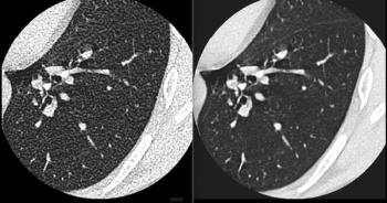
Contrast-enhanced multislice CT angiography, for example, is increasingly being used for the detection of coronary artery disease.11 Figure 5 shows an example of this type of imaging. The patient has a focal mixed calcified plaque in the left main coronary artery that was interpreted as a 50% to 60% stenosis. No other plaque was observed in this 67-year-old male, and his overall calcium score was at the 50th percentile level.
This patient was referred for CTA because of a mildly positive stress echocardiogram. His history was significant for chronic renal insufficiency. In this borderline situation, a radiologist, whose end-product is the official written report, would be understandably hesitant to take a clear position on whether the image data justify proceeding to invasive x-ray angiography with iodinated contrast, which might deteriorate renal function.
A cardiologist, conversely, must make a decision before going home at the end of the work day. In this circumstance, an official interpretation of "50% to 60% stenosis" would not be helpful. Convenient access to the images of Figure 4 would represent a major advantage, because the cardiologist could combine the raw image data with other information about the patient such as results of the most recent physical examination and the success of previous treatment strategies, considerably increasing confidence in the overall decision of how best to proceed. What the cardiologist really wants in this case is the ability to view the CTA side by side with the stress echocardiogram in order to quickly assimilate all of the relevant diagnostic information.
Cardiovascular disease remains the leading cause of death in the U.S., and diagnostic imaging is widely used to guide its treatment. To the cardiologist, viewing the results of imaging tests, particularly for borderline cases, is the primary reason for ordering them. Yet even today the only practical mechanism most cardiologists have to view these images is to walk to the scanner and hope someone will help.
No wonder cardiologists have "stolen" interventional angiography, echocardiography, and cardiac SPECT. Will this historic evolution happen again to MRI and CT, or will the equation be fundamentally altered by the emergence of EMRs and cardiac PACS? Clearly, only time can answer that question, but early recognition of the fundamental importance of remote image access presents an opportunity to influence the course of cardiac diagnostic imaging.
References
1. Transforming health care: the president's health information technology plan.
2. Brailer DJ, Thompson DG. The decade of health information technology: delivering consumer-centric and information-rich health care. U.S. Department of Health and Human Services. July 21, 2004.
3. Health Insurance Portability and Accountability Act (HIPAA). U.S. Department of Health and Human Services.
4. American College of Cardiology Cardiac Catheterization Committee. Cardiac angiography without cine film: erecting a "Tower of Babel" in the cardiac catheterization laboratory. J Am Coll Cardiol 1994;24(3):834-837.
5. Firefox: a free open-source Web browser.
6. Netscape: a free Web browser.
7. The World Wide Web Consortium (W3C). www.w3c.org.
8. Klem I, Shah DJ, Sketch MH Jr, et al. Improved detection of coronary artery disease by stress perfusion cardiovascular magnetic resonance with the use of delayed enhancement infarction imaging. J Am Coll Cardiol in press.
9. Simonetti OP, Kim RJ, Fieno DS, et al. An improved MR imaging technique for the visualization of myocardial infarction. Radiology 2001;218:215-223.
10. Kim RJ, Wu E, Rafael A, et al. The use of contrast-enhanced magnetic resonance imaging to identify reversible myocardial dysfunction. NEJM 2000;343:1445-1453.
11. Hoffmann MH, Shi H, Schmitz BL, et al. Noninvasive coronary angiography with multislice computed tomography. JAMA 2005:293(20):2471-2478.
Dr. Judd and Dr. Kim are codirectors, and Dr. Albert is a cardiology fellow, all at the Duke Cardiovascular Magnetic Resonance Center in Durham, NC. Mr. Umbach is a senior programmer at SciMed Solutions in Durham. Dr. Grizzard leads the cardiac imaging program at Virginia Commonwealth University in Richmond. Dr. Judd and Dr. Kim have an equity interest in Heart Imaging Technologies, which markets PACS and owns a related U.S. patent. Mr. Umbach is an employee of SciMed Solutions, which provides software products and information technology services to the healthcare and higher education markets.
CARDIAC PACS: CLICK BY CLICK
1 Click to load patient images
2. Multiple clicks on menu to learn how to advance to next series
3. Multiple clicks to select an image series
4. Multiple clicks on menu to learn how to open movie tool
5. Click to open movie tool
6. Click to play movie
7. Multiple clicks on menu to learn how control movie direction
8. Click to set movie speed
9. Multiple clicks to learn that movie speed cannot be set to patient's physiologic heart rate (not recorded by scanner)
10. Multiple clicks to learn that two series cannot be displayed side by side
Advertisement
Related Content
Advertisement


Advertisement
Advertisement
Trending on Diagnostic Imaging
1
FDA Clears AI-Powered Software for Improving Low-Contrast CT Detection
2
DeepHealth Launches AI-Powered Software for Radiology Reporting
3
Mammography Study: Can AI Detect Potential Breast Cancer Up to a Decade Prior to Diagnosis?
4
Thirteen Takeaways from New Report on AI in Health Care
5