Controlling Dose in the Emergency Department
Three tips to help you limit dose exposure and optimize imaging protocols.
Over the past decade radiation dose exposure has fallen precipitously. But, when you are asked to make split-second decisions in the emergency room, it can sometimes still be challenging to make the choices that will limit exposures.
In his presentation during the American Society of Emergency Radiology 2020 annual meeting, Mahadevappa Mahesh, M.S., Ph.D., chief physicist at The Johns Hopkins Hospital and professor of radiology and radiological sciences at Johns Hopkins Medicine, outlined several steps emergency radiologists can take to ensure you are controlling dose for your patients.
“We want to make sure that the people who are ordering the imaging understand the risks and benefits of imaging,” he told Diagnostic Imaging.
For more coverage of the ASER 2020 Annual Meeting, click
Since 2010, Mahesh said, CT dose exposure has fallen by nearly 20 percent. The use of more advanced medical imaging technologies and greater dose awareness campaigns can largely be credited for this success, as can provider education around optimizing imaging protocols. These are all critical achievements, he said, because in the emergency room, imaging should never be declined or denied with the clinical appropriateness is very high.
“One should not hesitate to do medical imaging, such as CT, X-ray, or fluoroscopy because they bring a lot of value,” he said. “But, in the emergency room, there is always a concern when we are ordering large imaging studies.”
Consequently, Mahesh shared several tips you can use to ensure that you are controlling exposure and optimizing protocols.
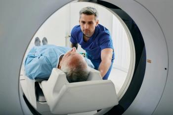
Have a Good Team: For those “in-the-moment” decisions about how much radiation dose you should really use with a patient in an emergency situation, Mahesh said, you must be sure you have the right people on your team. In addition to you and your colleagues, but sure to include two other individuals – a medical physicist and a technologist who reviews the protocols on an annual basis to ensure you’re using the right protocols. This team should also adapt all dose optimization strategies on the protocol. This is especially important with CT, he said, as they must pay attention to using modulation techniques.
Track Scan Utilization: As much as possible, keep tabs on the scans you are conducting. Monitor whether you are performing too many scans or repeating scans. Doing so will help you control utilization when – and if – it is necessary, he said.
Forego Lead Aprons: Although using a lead apron with patients has long been considered a protective strategy against radiation exposure, that paradigm is shifting, Mahesh said, particularly with gonad shielding in CT and X-ray. Foregoing their use can help you avoid duplicate studies.
“We are now advocating to stay away from placing the shield on the during imaging,” he said. “The American Association of Physicists in Medicine came up with a position statement that it is now advocating that it is not necessary to use the lead shielding – in fact, it may even be contraindicated. It can actually obscure objects in the path of the beam.”
In addition, he said, the lead apron can get shifted around, potentially interfering with the image quality, forcing you to repeat an image.
Overall, Mahesh said, you as the radiologists should concentrate efforts on being sure you and your colleagues are the providers who convey information to patients about radiation dose.
“The radiation risks should be explained or communicated by people who are knowledgeable about both the benefits and the risks of the imaging,” he explained.
For more coverage based on industry expert insights and research, subscribe to the Diagnostic Imaging e-newsletter
Newsletter
Stay at the forefront of radiology with the Diagnostic Imaging newsletter, delivering the latest news, clinical insights, and imaging advancements for today’s radiologists.


