
|Articles|October 24, 2005
CT body perfusion packages promise insights into diagnosis and therapy
Software from GE Healthcare and Siemens Medical Solutions is extending CT perfusion techniques beyond stroke assessment. Primary applications are in oncology, but much more may be possible.
Advertisement
Software from GE Healthcare and Siemens Medical Solutions is extending CT perfusion techniques beyond stroke assessment. Primary applications are in oncology, but much more may be possible.
Perfusion 3 from GE and syngo Body Perfusion-CT from Siemens are designed to evaluate perfusion of organs and tumors throughout the body, documenting changes in blood flow that may help differentiate between healthy and diseased tissue as well as monitoring the effects of therapy.
"Body perfusion offers a way of potentially quantifying the process of angiogenesis as a diagnostic indicator and determining the reaction of tumor tissue under treatment earlier than traditional CT, where the only thing you can measure is tumor volume changes," said Bernd Ohnesorge, Siemens' vice president of CT.
The two companies' packages are designed to assess tumors and other disease processes outside the brain, in the liver, bladder, kidney, or breast. The software has the potential to characterize tumors as benign or malignant by analyzing differences in perfusion parameters. GE reports users of its Perfusion 3 package differentiating between pancreatitis and pancreatic cancer.
Eventually, perfusion data may reduce or eliminate the need for tissue biopsies. At present, these analyses are only adjuncts to conventional means.
Siemens' syngo Body Perfusion-CT, released in early summer, calculates blood flow, blood volume, and vessel permeability from images reconstructed from dynamic CT data acquired after the injection of contrast media.
GE's Perfusion 3, released more than two years ago, allows body perfusion but particularly optimizes liver analyses. (GE's Perfusion 1 package focused only on the brain. Perfusion 2 extended studies to tumors in the brain and elsewhere.) The liver algorithms built into the latest product are particularly sophisticated, according to Karen Procknow, GE product development specialist for functional imaging.
"In the liver protocol, we have a number of different maps, such as the hepatic arterial fraction that tells you the percentage of the liver that is supplied by the hepatic artery versus the portal vein," she said. "It also calculates the time to enhancement, which shows the first point of enhancement across the liver."
Siemens Body Perfusion-CT is similarly advanced. The software allows the calculation of arterial and portal venous components, for example, while supporting the evaluation of regions of interest and the visual inspection of time density curves.
The latest perfusion software from GE and Siemens has potential for tracking patient response to therapy as well. Changes in parameters compared with baseline studies may indicate a positive or negative reaction, interpreted under specific clinical circumstances.
Output from the CT software includes images and data tables indicating flow rates, vessel permeability, and other parameters that indicate perfusion. Studies are typically conducted on high-performance CT scanners, such as Siemens Sensation 64 and GE's 64-slice LightSpeed VCT. Coverage afforded by the width of the detector is more important than speed, according to Procknow.
"It determines how much of the organ you can acquire," she said. "We do perfusion scans as cine or continuous acquisitions, which are then broken down into temporal resolution to map the perfusion."
In body perfusion, a specific volume, defined by the target organ or suspicious lesion, is scanned continuously to note differences in contrast enhancement. Gastric processes and respiration present challenges, as they may cause the tissue to move. Respiratory artifacts are kept in check mostly by scanning during breath-hold. Anatomic mapping algorithms detect and ameliorate remaining artifacts, Ohnesorge said.
Users apply dynamic scan protocols, then postprocess the data on advanced workstations such as Siemens' Leonardo and GE's Advantage Workstation.
The Siemens and GE body products sell for between $30,000 and $40,000. Perfusion 3, featuring brain and body capabilities as well as advanced liver analyses, lists for around $70,000.
Siemens reports strong interest from countries in Asia, where liver cancer is a major cause of death and physicians have limited access to PET and SPECT.
"Due to the very wide distribution of CT scanners, particularly in Asia, these countries are looking at the potential of this for adding information when doing tumor imaging," Ohnesorge said.
The potential of CT body perfusion is better known than its practicality, as early adopters are only beginning to document its clinical benefits. This dearth of documentation can be challenging in marketing these products.
"It is difficult in marketing, because there is very little we can say until the research proves it," said Bob Beckett, GE global product manager of diagnostic applications for functional imaging. "We are helping them look at perfusion changes, which may have different characteristics in inflammatory disease than in tumor."
As the technology's value in tumor assessment is better understood, the algorithms might be adapted for other applications. One possibility is CT perfusion of the heart, according to Ohnesorge.
"This is an emerging application," he said. "There is the potential to document ischemic processes."
Dynamic CT studies already are demonstrating perfusion defects, which appear as hypointense areas in the myocardium, according to Ohnesorge. Translating this fundamental data into clinical analyses would require segmentation algorithms to isolate the myocardium and color-code blood volumes in the myocardium. These data might then be interpreted in the context of other CT-derived information, such as the presence of stenosis.
To succeed, CT might have to follow the footsteps of nuclear cardiology, performing enhancement studies under rest and stress conditions. This may be possible with the latest generation of CT scanners, Ohnesorge said.
"Until recently, CT did not have the temporal resolution to scan patients at high heart rates," he said. "The 64-slice scanners have changed that."
Advertisement
Related Content
Advertisement


Advertisement
Advertisement
Trending on Diagnostic Imaging
1
SNMMI: What Early Research Reveals About the Alpha-Emitting Radioconjugate ATNM-400 for Prostate Cancer
2
Mammography Study Shows Impact of AI-Powered Slab Reconstruction with DBT
3
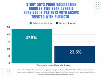
SNMMI: Does Vaccination Enhance Pluvicto Efficacy in mCRPC?
4
SNMMI: Over 80 Percent of Patients with Osseous Oligometastatic PCa are Upstaged with PSMA PET/CT
5