
|Articles|March 4, 2005
CT slice battle obscures value of flying focal spots
Siemens calls it z-Sharp. A competitor calls it z-Wobble. The names are ammunition in the battle for market share, underscoring the intensity of the fight more than they illuminate the technology.
Advertisement
Siemens calls it z-Sharp. A competitor calls it z-Wobble. The names are ammunition in the battle for market share, underscoring the intensity of the fight more than they illuminate the technology.
Siemens' z-Sharp, the cornerstone of the company's Sensation 64 CT scanner, electromagnetically directs fluxes of x-rays to their targets. Each pulse is fired moments apart, two through each point along the circumference of the gantry, multiplying the data obtained from each row on Siemens' solid-state detector by a factor of two. With this technology, 64 slices are pulled from a 32-row detector in each rotation, and 40 from a 20-row detector.
Siemens has used z-Sharp to take the lead among developers of 64-slice scanners. By the beginning of 2005, it had installed more than 100 such scanners at sites around the world. Its Sensation 64 has caught fire partly because competitors have been slow coming off the blocks. In January, Toshiba had installed fewer than a dozen of its Aquilion 64 systems. GE and Philips each had just one of their flagship 64-slice scanners operating at luminary sites.
But Siemens contends there is more to its success than being first to market. The technology delivers on its promise of improved resolution and coverage, according to the company. Customers on both sides of the Atlantic agree.
"I have the impression of coming from an ophthalmologist with new glasses," said Dr. Jonathan Kirsch, head of radiology at the Clinique Notre Dame de Tournai in Belgium. "The images are much sharper than before."
Siemens' 64-channel technology excels at vascular imaging, especially coronary CT angiography. Visualization of the vessel wall and its contents is clearly superior compared with previous platforms, said Dr. Jonathan Goldin, chief of radiology at the University of California, Los Angeles.
"This is partly because of the increased number of detectors, partly because of the engineering of the focal spot, and partly because of the rotational speed of the machine," Goldin said. "It's this combination that has led to improved temporal and spatial resolution."
Sensation 64 can complete a heart scan in eight seconds, accomplishing 0.4-mm resolution at 330-millisec rotation speed. The 0.4-mm spatial resolution can visualize a septal artery less than 1.5 mm in diameter and define plaque in the coronary arteries.
The system can conduct a gated chest exam in 20 seconds with resolution sharp enough to visualize a pulmonary aneurysm. Carotid stenoses demonstrating soft and calcified plaques can be seen in a six-second scan covering 27 cm from midskull to clavicle.
The improved resolution translates into better multiplanar and 3D reconstructions, according to Kirsch, who credits improvement in the z axis for better resolution. Kirsch estimates typical resolution at 0.4 mm versus 0.7 mm on a conventional 16-slice scanner.
"Now we can really work in 3D with all examinations. We can discover lesions that would otherwise be visible only in one or two planes," he said.
He referred to a case of a five-year-old trauma victim whose renal fracture was visible in the coronal and sagittal planes only.
The micromanagement of x-ray fluxes permits dose reductions, Goldin said. It also minimizes artifacts, according to Bernd Ohnesoge, vice president of global marketing and sales for Siemens CT. A Sensation 64 postoperative evaluation of a wrist and titanium implant produces clear bone details without the artifacts that otherwise accompany the presence of metal.
Siemens has doubled the number of detectors from 16 to 32 and doubled the amount of information obtained at each anatomic point with z-Sharp, Ohnesoge said.
"We have invested in a more clinically oriented concept, acquiring more data per rotation," he said. "Improved resolution and the reduction of artifacts become viable because of the way we do 64 slices."
Siemens' competitors do not address its claims of improved resolution or better image quality, focusing instead on the detector. GE describes the Sensation 64 as a 32-slice machine, citing Siemens' use of a 32-row detector. Building a 64-row detector is far more difficult than building one with 32, according to Siemens' competitors.
Ohnesoge dismisses the implication that Siemens' engineering accomplishments are less than those of its competitors because its premium detector has 32 rather than 64 rows.
"The quality of the detector shouldn't be measured by how many cuts are on its surface," he said. "Our electronics read out the data in a much more complex way than conventionally designed detectors."
Ohnesoge describes Siemens' Straton x-ray tube, the enabling technology behind z-Sharp, as a massive step forward in tube technology. Its electromagnetic guidance system serves essentially as a miniaturized electron-beam tomography system of the kind developed by Imatron, which was acquired by GE, and built into GE's e-Speed EBT cardiology system. The tube's cooling system eliminates the need for heat storage, allowing unlimited run times.
"The overall complexity and engineering challenges that went into our technology are at least as challenging as generating 64 slices the conventional way," he said. "We are proud of our innovation, but we're also proud that it translates into clinical benefits: higher resolution, better image quality, and dose minimization."
Siemens hopes to blaze a new trail with this technology. Its z-Sharp collimates the x-ray beam electromagnetically to 0.33 mm, rather than spreading it over the width of the detector row. The increased amount of data improves resolution, reducing or eliminating artifacts that occur around structures that strongly attenuate x-rays.
"These artifacts originate at high speeds from not sampling the data accurately enough," Ohnesoge said. "Our technology eliminates them by doubling the density of the data."
Siemens engineers are adapting z-Sharp to deliver x-rays at two different energies. Dual-energy CT promises to enhance contrast resolution, which may be especially helpful in vascular studies. The concept has been around for at least 20 years, according to Ohnesoge. It has been impractical because conventional CTs require two separate scans to acquire data sets at different energy levels, and the time elapsed between the scans makes coregistration of data all but impossible. Siemens' z-Sharp solves that problem.
"There are some very old concepts that were great ideas but were ahead of their time and now can be looked into," he said. "Who knows? Maybe the future of CT is in the history books."
Advertisement
Related Content
Advertisement


Advertisement
Advertisement
Trending on Diagnostic Imaging
1
SNMMI: What Early Research Reveals About the Alpha-Emitting Radioconjugate ATNM-400 for Prostate Cancer
2
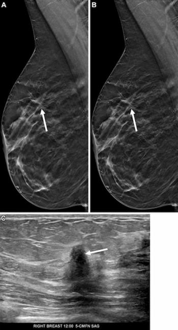
Mammography Study Shows Impact of AI-Powered Slab Reconstruction with DBT
3
SNMMI: Does Vaccination Enhance Pluvicto Efficacy in mCRPC?
4
GE HealthCare Unveils 4D Dynamic PET Imaging Software at SNMMI Conference
5