
|Articles|November 1, 2007
Digital mammography produces large data loads
Information management for digital mammography is unlike that of any other imaging modality. It generates huge files and enormous amounts of imaging data, which must then be stored, transmitted, and displayed.
Advertisement
Information management for digital mammography is unlike that of any other imaging modality. It generates huge files and enormous amounts of imaging data, which must then be stored, transmitted, and displayed.
While some may equate the size of imaging files from digital mammography with those produced using multislice CTs of the abdomen and pelvis, digital mammography is at a special disadvantage because it cannot make use of lossy compression. CT scans may be compressed on a 12:1 basis, on average, so they can be reduced to one-12th the size of the images in the original exam.
Lossy decompression admittedly loses some imaging data, but not much, said Dr. Michael Trambert, lead radiologist for PACS reengineering at Cottage Health System and the Sansum-Santa Barbara Medical Foundation Clinic. In fact, clinical studies have concluded that lossy decompression does not affect diagnostic quality or accuracy.
Many institutions around the world, however, do not employ lossy algorithms in mammography for legal reasons, at least not ones that produce the 12:1 space savings seen in CT. The U.S. Food and Drug Administration is particularly opposed to the use of such algorithms. But modern computing and networking, in combination with efficiencies achieved through staff management, have gone a long way toward making the argument for lossy compression moot.
Dr. Jean-Charles Piguet, a radiologist in the women's center at Imagerie & Development (ID) in Geneva, Switzerland, reports that standard networking at the privately run center simultaneously-and expeditiously-routes diagnostic mammograms obtained on two full-field digital mammography (FFDM) units to a workstation for soft-copy reading, hard-copy printing (a necessity for medicolegal reasons), and the PACS for storage.
Dr. Boel Heddson and colleagues at the Helsingborg Hospital in Helsingborg, Sweden, have similar success using a PACS to handle digital mammograms. She estimates wait time for soft-read digital mammograms in seconds. For her team, any longer would be too long.
Utilizing one FFDM system from Sectra and a computed radiography system from Fuji, the Swedish radiologists double-read about 110 screening mammograms and 15 to 20 clinical mammograms per day.
"We have a good RIS and PACS, working together, synchronized completely," Heddson said. "It goes fast."
This was not always the case, however. When Sansum-Santa Barbara Medical Foundation Clinic first introduced digital mammography in 2003, it had only a 100-MB mammography network. Radiologists consequently had to wait between 10 and 35 minutes to gain access to a mammographic study. A new 1-GB network has shaved access time to two to 10 minutes, Trambert said. Even a standard network fares well, such as the 500-MB one in use at the ID women's center, which sends images to print and soft copy simultaneously.
"As soon as you go out of the (examination) room to the workstation, it is already there," Piguet said. "It is not a problem; maybe 20 seconds pass; you don't notice it even."
The ease with which mammograms move from place to place within a healthcare system relies on the interconnectivity or bandwidth of the information management network, said Dr. Laurie Fajardo, a professor and chair of the radiology department at the University of Iowa in Iowa City.
"Networks can be of different sizes, and they can be composed of different components," she said. "They can get bogged down and create bottlenecks."
But the actual time that radiologists must wait between cases is negligible, if information technology administrators do their jobs well. The RIS at the ID women's center in Geneva is programmed to instruct the PACS to automatically prefetch past studies and send the data to both FFDM systems before the patient enters the examination room. Why both machines?
"Because we do not know which one the patient will be examined on," Piguet said.
Covering both makes past and just taken studies available at the soft-copy reading station with a mouse click.
"It's very convenient and easy," he said. "You just select the patient, open the file, and you have the images in two or three seconds."
But the huge files rendered by digital mammography can present challenges, if not in transmission, then in storage. Some breast centers save two sets of data, one composed of the so-called raw unprocessed data and the other full of processed data ready for display. Such a scheme can double or even triple the space needed for storage, according to Fajardo.
The ID women's center saves both sets because Piguet and colleagues are helping iCAD, a developer of computer-aided diagnostics, develop its mammography computer-aided detection for the Sectra digital mammography machine operating there.
Saving both sets is not uncommon, however, even among groups not involved in R&D. Some imaging facilities in the U.S. routinely save raw data from digital mammography so they can use CAD to follow the progression of suspicious areas from one visit to the next. This is necessary because CAD algorithms require raw data to find the suspicious lesions they highlight.
"When a woman comes back to have her mammogram the following year, if you don't have the for-processing data, you can't run her scans through CAD and compare what CAD saw last year and what it is seeing this year. If you get a more updated CAD algorithm, you can't run last year's mammograms through it unless you have the for-processing data," Fajardo said.
Imaging facilities around the globe also save raw data because of concerns about defensive medicine.
"Everyone is afraid to throw anything away. They're afraid of losing a tiny piece of information that might result in an error," Fajardo said.
Altogether, these storage demands can mount a substantial challenge to a PACS. How big a challenge depends on the size of the data set acquired, which varies substantially with the FFDM system. Some produce images as small as 8.4 MB. Others generate files as large as 53.5 MBs.
A study of U.S. imaging centers conducted by Fajardo indicates that a typical breast study stored using 4:1 lossless compression requires 8 to 15 MB of storage for a system rendering 100-micron resolution; 16 to 38 MB for a 70-micron system; and 45 to 60 MB for a 50-micron pixel detector imaging system.
Fajardo has calculated the image storage requirements for breast imaging centers with small, medium, and large patient volumes. Depending on the size of the pixel detector, centers that perform 7000 small-field-of-view and 3000 large-field-of-view exams in a year will need between 99 and 415 GB a year. The table shows storage needs depending on annual throughput.
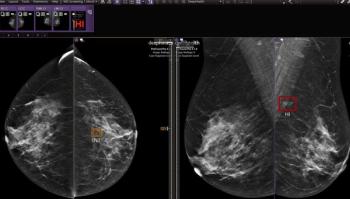
Proper planning can alleviate many of the concerns for both storage and transmission, just as efficient workflow alleviates bottlenecks. But even so, the travails of digital mammography are not necessarily over. The characteristically large data sets, particularly those comprising high-resolution images with 50-micron pixels, are too large to be viewed within the boundaries of 5-MP monitors. In such cases, only about 25% of a large breast might be seen in one view, according to Trambert.
Physicians consequently have to pan through the image section by section to see all the tissue. In the process, they may become confused about where they are looking and unable to compare densities in the contralateral breast. There may be challenges in just keeping the different images straight. Digital workflow scenarios may involve as many as 12 views, including all four new and old views, new and old craniocaudal views, new craniocaudal views on both screens, and new craniocaudal views in full resolution.
Engineers have responded with software fixes that allow the operator to shuffle through different images or arrange the images in a stack that can be scrolled back or forward. They may also use "mirrored panning," which allows radiologists to review images in full resolution, even if they extend off the monitor, as the viewer pans across the image, drawing in new tissue as old tissue exits the view screen.
Other PACS features, such as voice recognition and template-structured reporting, improve productivity for radiologists while providing referring physicians with a standardized format for reading the results of mammographic interpretations.
"A couple of clicks of the mouse make the whole workflow efficient for everyone," Trambert said.
Advertisement
Related Content
Advertisement


Advertisement
Advertisement
Trending on Diagnostic Imaging
1
What Does the Future Hold for Nuclear Medicine?
2
Top Five Radiology Content in June 2026
3
What New MRI Research Reveals About Endometrial Cancer Staging and the 2023 FIGO Staging System
4
FDA Clears New CEM Image Reconstruction and Biopsy Capabilities for Siemens Healthineers Mammography Device
5