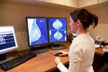
False-Positive Mammogram May Indicate Higher Breast Cancer Risk
False-positive screening mammograms may correspond with a higher risk of developing breast cancer.
Women with false-positive screening mammograms have a higher risk of a breast cancer diagnosis later on, according to a study published in
Researchers from Spain performed a retrospective cohort study to assess the risk of breast cancer in women who had received false-positive screening results, according to radiologic classification of the features seen on mammography.
The cohort study included 521,200 women (aged 50 to 69), who underwent screening as part of the Spanish Breast Cancer Screening Program between 1994 and 2010 and who were observed until December 2012. False-positive results were defined as a screening mammogram with a BI-RADS score of 3, 4, 5, or 0. A total of 76,075 women in this cohort had false-positive results.[[{"type":"media","view_mode":"media_crop","fid":"46754","attributes":{"alt":"mammography","class":"media-image media-image-right","id":"media_crop_7988900435941","media_crop_h":"0","media_crop_image_style":"-1","media_crop_instance":"5444","media_crop_rotate":"0","media_crop_scale_h":"0","media_crop_scale_w":"0","media_crop_w":"0","media_crop_x":"0","media_crop_y":"0","style":"height: 120px; width: 180px; border-width: 0px; border-style: solid; margin: 1px; float: right;","title":"©zlikovec/Shutterstock.com","typeof":"foaf:Image"}}]]
The results showed that the age-adjusted hazard ratio (HR) of women with false-positive results was 1.84, compared with those women who had negative mammograms. The risk was higher among women who had calcifications, whether the calcifications were associated with masses or not. “Women in whom mammographic features showed changes in subsequent false-positive results were those who had the highest risk,” the authors wrote.
“Women who had more than one examination with false-positive findings and in whom the mammographic features changed over time had a highly increased risk of breast cancer,” they concluded. “Previous mammographic features might yield useful information for further risk-prediction models and personalized follow-up screening protocols.”
Newsletter
Stay at the forefront of radiology with the Diagnostic Imaging newsletter, delivering the latest news, clinical insights, and imaging advancements for today’s radiologists.


