FDA Grants Expanded Approval of Siemens Healthineers Mammography Platform for Digital Breast Tomosynthesis
The inclusion of the 3D imaging technology PlatinumTomo in the Mammomat B.brilliant device reportedly enables clinicians to obtain 50-degree 3D images in less than five seconds.
Offering a variety of new features for improved efficiency and enhanced 3D breast imaging, the mammography platform Mammomat B.brilliant has garnered an expanded approval from the Food and Drug Administration (FDA).
Building upon the 50-degree wide-angle technology of its existing 3D mammography systems, Siemens Healthineers says the expanded approval for the device includes PlatinumTomo, which reduces blurring that commonly occurs with 3D imaging and allows one to obtain 50-degree 3D images in less than five seconds.


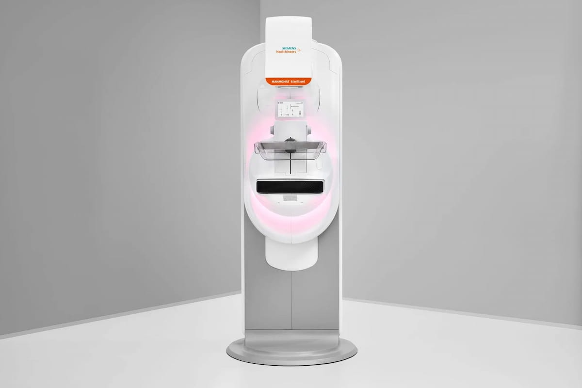
New features with the Mammomat B.brilliant platform include PlatinumTomo, which enables five-second acquisition of 50-degree 3D images, and UltraHD technology, which reduces metal artifacts and provides enhanced visualization of calcifications, according to Siemens Healthineers, the manufacturer of the device. (Image courtesy of Siemens Healthineers.)
Other new features with the Mammomat B.brilliant platform include a new X-ray tube, which includes adaptation of the flying focal spot technology from the company’s computed tomography (CT) scanners, according to Siemens Healthineers, the manufacturer of the Mammomat B.brilliant Device.
The company pointed out that the expanded approval also includes UltraHD technology for image reconstruction that facilitates enhanced calcification visualization, reduced metal artifacts and the availability of synthetic 2D images without exposing patients to additional radiation.
“This revolutionary system not only provides health-care institutions with significantly improved diagnostic capabilities but also addresses the critical need for patient and technologist comfort in breast cancer screening,” added Niral Patel, the vice president of X-ray products at Siemens Healthineers North America.
Newsletter
Stay at the forefront of radiology with the Diagnostic Imaging newsletter, delivering the latest news, clinical insights, and imaging advancements for today’s radiologists.


Mammography Study: AI Facilitates Greater Accuracy and Longer Fixation Time on Suspicious Areas
July 8th 2025While noting no differences in sensitivity, specificity or reading time with adjunctive AI for mammography screening, the authors of a new study noted a 4 percent higher AUC and increased fixation time on lesion regions.


Can Contrast-Enhanced Mammography be a Viable Screening Alternative to Breast MRI?
June 17th 2025While the addition of contrast-enhanced mammography (CEM) to digital breast tomosynthesis (DBT) led to over a 13 percent increase in false positive cases, researchers also noted over double the cancer yield per 1,000 women in comparison to DBT alone.

Contrast-Enhanced Mammography and High-Concentration ICM Dosing: What a New Study Reveals
June 16th 2025New research showed a 96 to 97 percent sensitivity for contrast-enhanced mammography (CEM) with an increased iodine delivery rate facilitating robust contrast enhancement for women with aggressive breast cancer.

