GE Unveils Three New MRI Technologies at ISMRM
Products focus on Alzheimer’s disease, traumatic brain injury, and accelerating clinical translation.
GE Healthcare is showcasing three new technologies during this year’s International Society of Magnetic Resonance in Medicine virtual annual meeting. These new tools are dedicated to enhancing neurological research with Alzheimer’s disease and traumatic brain injury.
According to a company statement, GE is highlighting these new tools:
SIGNA 7.0T: This 510(k)-pending technology, the company said, has approximately five times more power than most clinical systems and is designed to pick up subtle clinically- and research-relevant structures It features a 60-cm bore system and is outfitted with UltraG gradient technology, GE’s most powerful, whole-body gradient coil.


Left quantified changes in the brain. Sample images obtained on SIGNA 7.0T system 510(K)-pending at FDA. Not available for sale. Courtesy: GE Healthcare
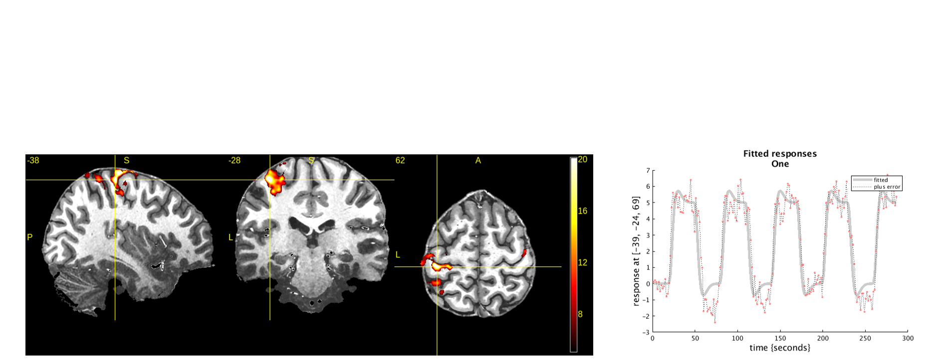
With Precision RF transmit-and-receive architecture, it can facilitate improved image quality and research flexibility, company officials said. Additionally, clinicians will be able to use the existing SIGNAWorks applications platform to access deep learning-based platform tools, such as AIR x brain and Silent MR imaging.
SIGNA UHP 3.0T: Similar in some ways to the SIGNA 7.0T, this investigational device is a whole-body 3T system that can scan all clinical anatomies, and, according to a company press release, it is designed for research and can provide improved visualization of microstructure changes associated with Alzheimer’s and Parkinson’s. Its lower field strength – and, therefore, lower cost – makes the research gains from its UltraG gradient technology, RF architecture, and SIGNAWorks features accessible to more institutions, company officials said.
MAGNUS: GE is also revealing its experimental high-performance Microstructure Anatomy Gradient for Neuroimaging with Ultrafast Scanning (MAGNUS). According to company data, the U.S. Department of Defense-funded technology will be installed in a U.S. military treatment facility to study new imaging biomarkers that can be used to diagnose mild traumatic brain injury.


MAGNUS images; sample imagines obtained on research device limited by U.S. law to investigational use. Courtesy: GE Healthcare
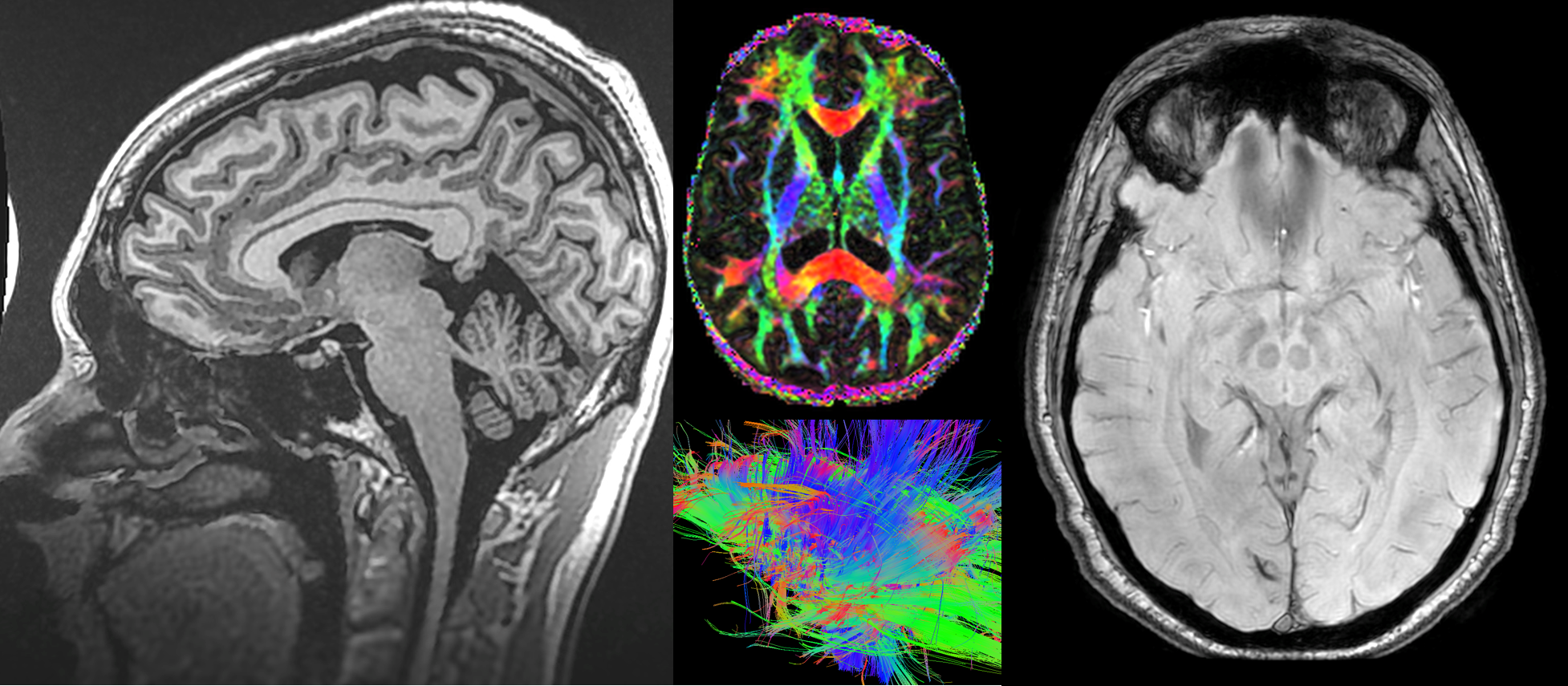
MAGNUS will have a different gradient strength and slew rate for brain imaging than conventional MRI scanners, the company said, operating in the 500-700T/m/s and 200-300 mT/m range rather than 200T/m/s and 50-80 mT/m. The goal, said Jie Xue, president and CEO of GE Healthcare MR, is to potentially establish the first objective measures for diagnosing mild traumatic brain injury cases, as well as Alzheimer’s, Post-Traumatic Stress Disorder, Attention Deficit Disorder, and mild cognitive impairment.
Newsletter
Stay at the forefront of radiology with the Diagnostic Imaging newsletter, delivering the latest news, clinical insights, and imaging advancements for today’s radiologists.


The Reading Room Podcast: Emerging Trends in the Radiology Workforce
February 11th 2022Richard Duszak, MD, and Mina Makary, MD, discuss a number of issues, ranging from demographic trends and NPRPs to physician burnout and medical student recruitment, that figure to impact the radiology workforce now and in the near future.


