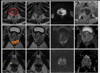
An AI model for prostate MRI offered an 89 percent AUC for prostate cancer detection in external validation testing, according to a newly published international multicenter study.
In a recent video interview, Amar Kishan, M.D., discussed a new study that demonstrated significant side effect reduction when utilizing magnetic resonance imaging (MRI) guidance instead of computed tomography (CT) guidance for stereotactic body radiation therapy (SBRT) to treat localized prostate cancer.
Emerging research suggests that magnetic resonance imaging (MRI) guidance may be more advantageous than computed tomography (CT) guidance for reducing the side effects of stereotactic body radiation therapy (SBRT) in the treatment of prostate cancer.
Results from the phase 3 randomized trial, recently published in
In a recent video interview, lead study author Amar Kishan, M.D. called the MRIdian LINAC (ViewRay) a “major technological breakthrough” that allows physicians to combine continuous MRI imaging with a more targeted radiation delivery system for
“(With) these advanced precision and accuracy capabilities, particularly that high degree of tracking during the treatment, we felt we could reduce the margins around the prostate that we need to treat from four millimeters to two millimeters,” explained Dr. Kishan, an associate professor, vice-chair of Clinical and Translational Research and chief of the Genitourinary Oncology Service within the Department of Radiation Oncology at UCLA.
For more insights from Dr. Kishan, watch the video below.