
|Articles|April 29, 2013
Integrated 2D and 3D Mammography Detects More Cancers
Author(s)Diagnostic Imaging Staff
Integrating 2D and 3D screening increases breast cancer detection and may lower reports of false positives.
Advertisement
Integrated 2D and 3D mammography improves breast-cancer detection and may reduce false positive recalls, according to a study published in
Conventional 2D mammography for breast cancer screening has limitations in both detecting cancer and producing false positive results. To address this issue, researchers from Italy and Australia investigated how integration of 2D and 3D mammography may improve cancer detection. This has been looked in a few smaller studies, but this is the largest completed trial that reported on the effectiveness of 3D screening in a larger group.
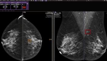
A total of 7,292 women, median age 58, were screened. Readings were first done with 2D imaging alone and then with integrated 2D and 3D mammography, resulting in paired data for each screening.
The researchers were looking at final assessment or excision histology. Primary outcome measures were the number of detected cancers, the number and proportion of false positive recalls, and incremental cancer detection attributable to integrated 2D and 3D mammography.
Fifty-nine cancers were detected in 57 patients: 20 cancers were found with integrated 2D and 3D versus none with 2D screening only. The cancer detection rates were 5.3 cancers per 1,000 screens when 2D alone was used and 8.1 cancers per 1,000 screens with integrated 2D and 3D screening. The incremental cancer detection rate attributable to integrated 2D and 3D mammography was 2.7 cancers per 1,000 screens.
False results were reported from a total of 395 screens: 181 at both screen reads, and 141 with 2D only, versus 73 with integrated 2D and 3D screening. The researchers estimated that conditional recall (positive integrated 2D and 3D mammography as a condition to recall) could have reduced false positive recalls by 17.2 percent without missing any of the cancers detected in the study population.
The researchers concluded that the integration of 2D and 3D mammography raised detection levels and that randomized controlled trials are needed for further investigation.
Advertisement
Related Content
Advertisement


Advertisement
Advertisement
Trending on Diagnostic Imaging
1
What Does the Future Hold for Nuclear Medicine?
2
Top Five Radiology Content in June 2026
3
What New MRI Research Reveals About Endometrial Cancer Staging and the 2023 FIGO Staging System
4
FDA Clears New CEM Image Reconstruction and Biopsy Capabilities for Siemens Healthineers Mammography Device
5