Key MRI Patterns for Diagnosing Inflammatory Brain Stem Lesions
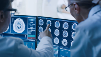
In an award-winning presentation at the European Congress of Radiology (ECR), researchers from the Hospital Universitario Basurto in Spain reviewed key neuroimaging findings on magnetic resonance imaging (MRI) for a variety of inflammatory lesions of the brain stem.
Noting the variety of disorders that can lead to inflammatory brain stem lesions, researchers discussed pertinent magnetic resonance imaging (MRI) findings for diagnosing the lesions in a poster presentation that earned magna cum laude honors at the European Congress of Radiology (ECR) in Vienna, Austria.1
Inflammatory lesions in the brain stem can be challenging to diagnose, according to poster co-author Juan Jose Gomez Muga, MD, who is affiliated with the Radiology Department at the Hospital Universitario Basurto in Spain, and colleagues.
“Some autoimmune, paraneoplastic, metabolic and infectious disorders may show similar appearances (with brain stem lesions), making an accurate approach difficult,” wrote Muga and colleagues.
Emphasizing key signs and patterns on MRI, the poster authors reviewed neuroimaging findings for 16 disorders, ranging from Neuro-Behcet disease and neurosarcoidosis to Bickerstaff brainstem encephalitis and systemic lupus erythematosus, that are among the most common etiologies for inflammatory brain stem lesions.
For neuromyelitis optica spectrum disorder (NMOSD), one would see mostly infratentorial lesions near the fourth ventricle. The linear, T2-hyperintense lesions may be contiguous with cervical transverse myelitis, according to the poster authors. They added that other key MRI findings include the appearance of diencephalic lesions in proximity to the third ventricle and rostral midbrain.
With Neuro-Behcet disease, Muga and colleagues said T2-weighted MRI will show edematous lesions that have a hyperintense appearance while one will note microhemorrhages on susceptibility-weighted imaging and contrast enhancement of acute lesions on T1-weighted MR images. Optic neuritis and intracranial sinus thrombosis are common with Neuro-Behcet diseases, according to the poster authors. Muga and colleagues also noted that infratentorial atrophy may result from chronic Neuro-Behcet disease.
When it comes to brain stem lesions caused by multiple sclerosis, Muga and colleagues said radiologists will see well-circumscribed, ovoid lesions that are found in the dorsal and ventral brain stem. More likely to occur in the pons than the medulla, these lesions are hyperintense on fluid-attenuated inversion recovery (FLAIR) sequences and T2-weighted MRI images, according to the poster authors.
Reference
1. De Andoin Sojo CG, Anton L, Sanchez LA, et al. Inflammatory lesions of the brainstem: MRI patterns to success in diagnosis. Poster presented at: European Congress of Radiology; July 13-17; Vienna, Austria.
Newsletter
Stay at the forefront of radiology with the Diagnostic Imaging newsletter, delivering the latest news, clinical insights, and imaging advancements for today’s radiologists.


