For patients who survived sudden cardiac arrest (SCA), cardiac magnetic resonance imaging (MRI) identified the correct diagnosis in more than a third of cases in which other modalities had inconclusive findings in one study and led to a changed diagnosis in 13 to 50 percent of cases across multiple studies, according to a new literature review.
For the systematic review, recently published in Radiology: Cardiothoracic Imaging, researchers reviewed data from 14 studies (published between 2012 and January 2023) and a total of 1,367 patients who had SCA. According to the review, 91.9 percent had cardiac MRI exams.
The researchers noted a variety of inconsistent findings on the diagnostic yield of cardiac MRI. While two studies reported an ischemic cardiomyopathy (ICM) incidence of less than 10 percent and two studies noted no cases of ICM, six studies reported an ICM incidence of 40 percent and higher. One 2021 study, involving 121 patients who received cardiac MRI, reported an ICM incidence of 62 percent.
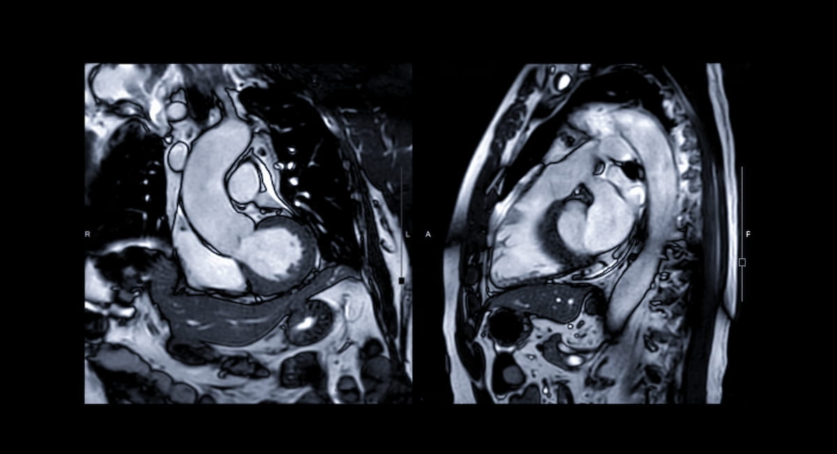
“ … Cardiac MRI proves invaluable in delineating regions of myocardial ischemia and infarction. The high spatial resolution of the technique allows for the identification of myocardial scar tissue and assessment of perfusion abnormalities, contributing substantially to the understanding of ischemic heart diseases,” wrote review co-author Reinhard Kaufmann, M.D., who is affiliated with the Department of Radiology at Paracelsus Medical University in Salzburg, Austria, and colleagues.
In regard to changed diagnoses, one study deemed cardiac MRI as “not useful” and another study concurred with no resulting diagnoses uncovered by the modality. However, two of the other reviewed studies noted the “crucial” nature of cardiac MRI in diagnosing 30 percent of cases and achieving correct diagnosis in 36.4 percent of cases in which findings from other modalities were non-diagnostic. Multiple studies found that cardiac MRI led to changed diagnoses that affected 13 to 50 percent of cases, according to the review authors.
“Cardiac MRI led to a new or alternate diagnosis in patients who survived SCA, secondarily influencing the management of patients,” added Kaufmann and colleagues.
Three Key Takeaways
- Diagnostic value of cardiac MRI. Cardiac MRI proved to be a valuable tool in diagnosing patients who survived sudden cardiac arrest (SCA), particularly when other modalities yielded inconclusive results. For one study, in more than a third of cases in which other methods were inconclusive, cardiac MRI identified the correct diagnosis. Moreover, across multiple studies, it led to a changed diagnosis in a significant percentage of cases, ranging from 13% to 50%.
- Impact in detecting ischemic cardiomyopathy (ICM). While some studies reported lower detection of ICM with cardiac MRI, nearly half of the reviewed studies noted an ICM incidence of 40 percent or higher with one of the studies reporting a 62 percent incidence.
- Complementary role with transthoracic echocardiography (TTE). While TTE remains the first-line imaging modality for assessing the cause of SCA, cardiac MRI provides superior detail in delineating myocardial texture and subepicardial structures. Combining both modalities may enhance clinical decision-making by offering a comprehensive view of cardiac structure, function, and tissue composition, thereby facilitating more informed diagnostic and therapeutic strategies for patients recovering from SCA.
While the review authors acknowledged that transthoracic echocardiography (TTE) is first-line imaging to ascertain the cause of SCA, they maintained that cardiac MRI provides superior detail with respect to myocardial texture and subepicardial structures. The researchers suggested that the combination of TTE and cardiac MRI could elevate the management of patients recovering from SCA.
“This combined approach enhances clinical decision-making and elevates the standard of patient care by providing a holistic view of cardiac structure, function, and tissue composition, thereby facilitating more informed diagnostic and therapeutic strategies for patients with various cardiomyopathies,” noted Kaufmann and colleagues.
(Editor’s note: For related content, see “Can Deep Learning MRI Have an Impact in Suspected Stroke Cases?,” “Seven Takeaways from New Consensus Recommendations for Cardiac MRI Assessment of COVID-19” and “Emerging AI Advances in Cardiac Imaging.”)
In regard to limitations with the review, the authors noted the lack of comparison between index cardiac MRI and other modalities such as TTE in 11 of the 14 studies reviewed. Variations with study cohorts — including studies focusing only on patients with SCA and other studies emphasizing cohorts of patients with non-ischemic cardiomyopathy (CMP) — also led to comparative challenges, according to the researchers. They added that the lack of randomization in most of the reviewed studies thwarted conclusive findings on the accuracy of cardiac MRI.