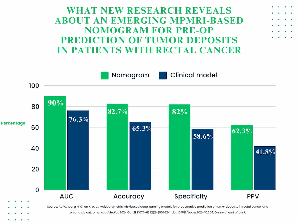
External validation testing of an emerging deep learning (DL) nomogram revealed significantly enhanced accuracy, specificity, and positive predictive value (PPV) in preoperative prediction of tumor deposits in patients being treated for rectal cancer.
For the retrospective study, recently published in Academic Radiology, researchers compared a clinical model (based on magnetic resonance imaging (MRI)-detected T stage and lymph nodes), a 30-feature DL model and a nomogram that combined DL and clinical factors for predicting tumor deposits in 529 patients who had radical surgery for rectal cancer.
In external validation testing, the nomogram demonstrated significantly higher capacity for predicting tumor deposits than the clinical model with respect to area under the curve (AUC) (90 percent vs. 76.3 percent), accuracy (82.7 percent vs. 65.3 percent), specificity (82 percent vs. 58.6 percent) and positive predictive value (PPV) (62.3 percent vs. 41.8 percent).
“One possible explanation is that (tumor deposit) is more prevalent in patients with locally advanced (rectal cancer), and deep learning features derived from such cases may show significant differences in radiomic characteristics compared to early-stage cases,” wrote lead study author Weiqun Ao, M.D., who is affiliated with the Department of Radiology at the Tongde Hospital of Zhejiang Province in Hangzhou, China, and colleagues.
While multiparametric MRI (mpMRI) plays a key role in defining tumor biology and prognostic evaluation of patients with rectal cancer, the researchers noted challenges with the heterogenous nature of perirectal fat lesions and overlap between the presentations of tumor deposits and non-tumor deposit lesions.
Three Key Takeaways
1. Enhanced prediction accuracy with deep learning (DL) nomogram. Combining MRI-based clinical factors with a 30-feature DL model, the nomogram significantly improved predictive accuracy (82.7 percent vs. 65.3 percent) and specificity (82 percent vs. 58.6 percent) over the clinical model in identifying tumor deposits in rectal cancer.
2. Prognostic value for disease-free survival (DFS). Both the DL model and nomogram successfully differentiated between high- and low-risk groups for three-year DFS, unlike the clinical model, indicating their potential in evaluating prognosis in patients with rectal cancer.
3. Potential impact on clinical decision-making. The DL model and nomogram demonstrated superior ability to identify patients at higher risk for recurrence, which could aid clinicians in making more informed treatment decisions. By accurately identifying high-risk patients, these tools may help personalize therapeutic strategies and improve outcomes in rectal cancer management.
Emphasizing that tumor deposits are often associated with lower survival in patients with rectal cancer, the study authors noted the clinical model failed to differentiate between patients at high- and low-risk for three-year disease-free survival (DFS) at one of the two participating centers in the study.
“In contrast, both the DL model and the nomogram successfully differentiated between high- and low-risk patients with distinct DFS profiles, demonstrating superior predictive capabilities,” pointed out Ao and colleagues. “Additionally, applying the DL model and the nomogram in predicting 3-year DFS outcomes supports their potential role in the prognostic evaluation of (rectal cancer).”
(Editor’s note: For related content, see “Rectal Cancer MRI: Seven Key Takeaways from a New Literature Review,” “Diffusion-Weighted MRI and Neoadjuvant Chemotherapy for Rectal Cancer: What New Research Reveals” and “Hybrid MRI Deep Learning Model Shows Promise in Predicting Tumor Deposits with Rectal Cancer.”)
In regard to study limitations, the authors acknowledged the possibility of patient selection bias given the study’s exclusion of patients who had preoperative neoadjuvant therapy. The researchers also noted the lack of assessment for tumoral and peritumoral features.
