Low-field magnetic resonance imaging (MRI) findings from a new study revealed that moderate to severe opacities in the lungs were a persistent finding for a significant number of patients up to two years after having acute COVID-19 pneumonia.
For the retrospective study, recently published in Academic Radiology, researchers reviewed data from 167 follow-up low-field MRI exams for 104 patients (mean age of 55) who had acute COVID-19 pneumonia. According to the study, the authors assessed MRI findings from five different timeframes in the study: within six months of infection (32 patients); between six to 12 months after infection (63 patients); between 12 and 18 months (42 patients); between 18 months and 24 months (24 patients); and beyond 24 months (six patients). The study authors noted 63 serial exams and that 32 patients had more than one MRI exam.
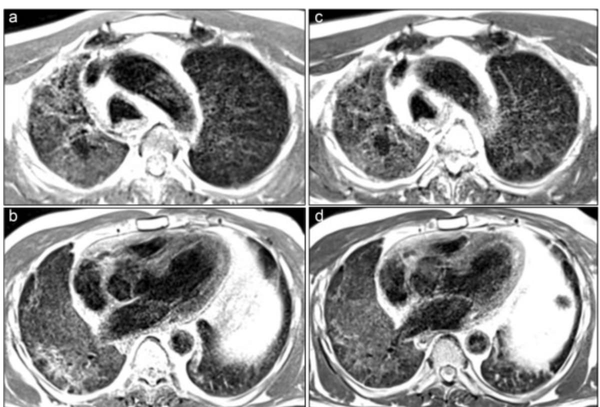
Using a quadrant grading system for assessing pulmonary findings, the study authors utilized opacity scoring for categories of no opacity (0), moderate opacity (>8), severe opacity (>12) and opacity>75 percent (16).
The researchers found moderate to severe opacities in a third of the exams conducted at more than six months after initial acute COVID-19. Moderate to severe opacities appeared on 25 percent of low-field MRI exams conducted between 12 to 18 months after illness and for three of the six patients who had follow-up MRI imaging beyond 24 months, according to the study authors.
For the 32 patients who had serial examinations, the researchers noted the prevalence of persistent opacity with 11 patients having no change in opacity score and 11 patients showing increased opacity scores from initial imaging to final examinations.
“ … Longitudinally persisting opacities were commonly identified by low-field MRI in post-acute COVID-19 patients, and initial disease severity was associated with increased MRI opacity score up to 24 months,” wrote lead study author Lea Azour, M.D., an associate professor of radiology at the David Geffen School of Medicine at UCLA, and colleagues.
Three Key Takeaways
1. Long-term lung opacities. The study indicates that moderate to severe opacities in the lungs persist in a significant number of patients up to two years after experiencing acute COVID-19 pneumonia. This suggests that there may be prolonged pulmonary effects associated with COVID-19.
2. Persistence and increases in opacity scores for majority of patients with serial imaging. Approximately 69 percent of patients who had serial low-field MRI follow-up exams had no change in opacity score or increases in opacity score from initial imaging to final imaging.
3. Potential of low-field MRI for lung surveillance. In addition to superior specificity and negative predictive value in comparison to CT, the authors said low-field MRI may offer enhanced parenchymal visualization and less susceptibility to artifacts at air-tissue interfaces.
Noting increased T1 signal intensity and less susceptibility to artifacts at air-tissue interfaces, the study authors suggested that low-field MRI (modified from 1.5 T MRI to operate at 0.55 T) may offer unique advantages as a radiation-free alternative for lung surveillance.
“The utility of MRI for surveillance imaging has been suggested at conventional field strengths, for example due to its superior specificity and negative predictive value than CT,” noted Azour and colleagues. “ … While conventional field strength MRI has been used for lung imaging including in cystic fibrosis and (interstitial lung disease) populations, low field imaging has potential to further improve parenchymal visualization. In addition, the lower siting and maintenance costs can increase patient access to this technology and broaden use of MRI for lung imaging.”
(Editor’s note: For related content, see “MRI Research Suggests Link Between COVID-19 Related Brain Fog and Blood-Brain Barrier Dysfunction,” “Can Diffusion Microstructural Imaging Provide Insights into Long COVID Beyond Conventional MRI?” and “MRI Study: People with Past Hospitalization for COVID-19 Have Nearly Triple the Risk of Multiorgan Abnormalities.”)
Beyond the inherent limitations of a retrospective study, the researchers acknowledged variability with the number of MRI exams obtained at different timepoints in the study and that assessment of opacity severity was limited to T2-weighted sequences.