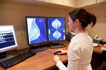
New management brings acoustical holography to market
The thrust for Richland, WA-based Advanced Imaging Technologies over the last four years has been to transform the acoustical holography system developed by its predecessor firm, Advanced Diagnostics, into a commercial product. The fruit of those labors is the company’s first product, its Aria Breast Imaging System.
The thrust for Richland, WA-based Advanced Imaging Technologies over the last four years has been to transform the acoustical holography system developed by its predecessor firm, Advanced Diagnostics, into a commercial product. The fruit of those labors is the company's first product, its Aria Breast Imaging System.
The product is the result of work performed by a management team headed by company president and CEO Lura J. Powell. Aria, shown at the 2006 RSNA meeting, takes advantage of the diffractive properties of ultrasound waves to obtain boundary and tissue information about soft structures, such as those in the breast. Aria focuses sound waves on a proprietary detector that makes more than 100 3D holograms a second. In the process, it can capture 97% of the sound waves that pass through the breast tissue, presenting anatomic data in multislice, multiplanar views.
"This is not something where you gather data and reconstruct what's inside the breast. What you see are 3D holograms of what is inside breast tissue, including ducts, ligaments, blood vessels, and cancerous and other types of lesions," Powell said.
Although AIT's acoustical holography technology has FDA clearances for use in pediatric, vascular, and large and small musculoskeletal imaging, the company is initially targeting the breast.
"The technology has general imaging capabilities, but as a company that is commercializing something new, we felt we had to focus, and breast imaging is where we believe we can do the most good right now," she said.
The company is concentrating on imaging dense breast tissue. On mammography screenings of dense breasts, particularly dense volumes and lesions appear in white, according to Powell. The Aria distinguishes between these two, depicting lesions as darker than dense masses, which absorb more sound. The ultrasound system also reveals spiculated capsules, irregular margins, and microlobulations in malignant masses. Cysts, which are less dense structures, appear light on Aria images, with smooth edges.
"About 30% to 40% of women have dense breast tissue, so it's important to have a technology that can effectively see through dense breasts and give points of reference in whole breast imaging so radiologists can quickly scan to localize any lesions and be sure they are not missing anything," Powell said.
Newsletter
Stay at the forefront of radiology with the Diagnostic Imaging newsletter, delivering the latest news, clinical insights, and imaging advancements for today’s radiologists.


