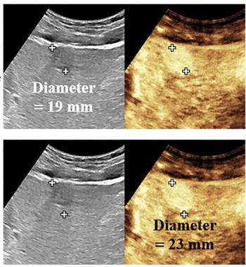
For Breast Imaging Reporting and Data System (BI-RADS) 4 cases, the study authors noted that in contrast to FFDM alone, FFDM/UST had non-inferior specificity (82 percent vs. 88 percent) but offered superior sensitivity (37 percent vs. 30 percent). However, in these cases, the researchers pointed out a net increase of 5.5 more false positive findings per reader.
“(The) addition of UST to FFDM resulted in only a slight reduction in positive predictive value compared with FFDM alone (42.2% vs 45.4%, respectively),” cautioned Yamashita and colleagues. “Clinical studies are needed to determine whether supplemental UST can improve cancer detection without substantially increasing the number of benign biopsies.”
While the researchers saw no statistically significant difference with mean sensitivity in BI-RADS 3 cases (40 percent for FFDM/UST vs. 33 percent for FFDM alone), they found that FFDM/UST provided superior mean specificity (75 percent vs. 69 percent).
“The specificity improvement with FFDM plus UST at BI-RADS 3 assessment is encouraging. A BI-RADS 3 assessment is the most common source of false-positive findings in handheld US and automated breast us screening,” noted Yamashita and colleagues. “With improved BI-RADS 3 specificity, radiologists can more accurately characterize masses as benign (BI-RADS 2), leading to fewer BI-RADS 3 assessments.”
Three Key Takeaways
1. Enhanced detection accuracy. Combining full-field digital mammography (FFDM) with whole-breast ultrasound tomography (UST) improves breast cancer detection accuracy in women with dense breasts. The combination yielded an area under the curve (AUC) of 60 percent, compared to 54 percent for FFDM alone.
2. Superior sensitivity in BI-RADS 4 cases. For BI-RADS 4 cases, FFDM combined with UST showed superior sensitivity (37 percent vs. 30 percent) and non-inferior specificity (82 percent vs. 88 percent) compared to FFDM alone. This suggests that adding UST can help identify more cancers that might be missed by mammography alone, although it comes with a slight increase in false positives.
3. Increased false positive findings. While the researchers noted the benefits of UST with respect to coronal volumetric image sequences and stiffness fusion sequences, the addition of UST to FFDM resulted in an increase in false positive findings. Specifically, there was a net increase of 5.5 more false positives per reader. This concern highlights the need for clinical studies to determine if the benefits of improved cancer detection outweigh the drawbacks of increased benign biopsies.
In an accompanying editorial, Ritse Mann, M.D., Ph.D, said the increased sensitivity with UST for BI-RADS 4 cases is in line with previous research looking at supplemental ultrasound but expressed reservation about the “poor overall sensitivity” and likelihood of increased false-positive findings.
“In my opinion, this study shows that the current implementation for UST is at best on par with handheld US and automated breast US, allowing detection of some additional cancers at the price of a substantial increase in the number of false-positive findings. It seems that UST may be a new implementation of US, but it ultimately yields old results,” maintained Dr. Mann, the chair of breast imaging at the Netherlands Cancer Institute in Amsterdam and chair of the scientific and program committees of the European Society of Breast Imaging.
(Editor’s note: For related content, see “Leading Breast Radiologists Discuss the USPSTF Breast Cancer Screening Recommendations,” “Whole Breast Ultrasound Screening: Is There Adequate Utilization in Patients at Higher Risk for Breast Cancer?” and “Breast Ultrasound Study: AI Radiomics Model May Help Predict Lymphovascular Invasion in Breast Cancer Cases.”
In regard to study limitations, the authors conceded that reviewing radiologists received abbreviated clinical histories, had no prior imaging for patients and interpreted images in a laboratory setting for the study. The researchers also pointed out that the reviewing radiologists had no prior experience with interpreting images from the breast ultrasound tomography (UST) system utilized in the study.