Next-generation advances push PET/CT into new clinical ground
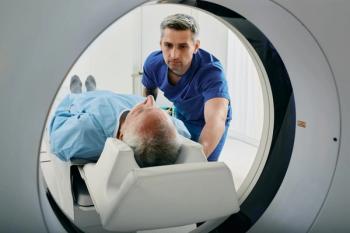
A new generation of hybrid scanners has entered the clinical mainstream. Featuring high-performance PET detectors and 16-slice CTs, these systems have followed their predecessors' path into oncology, but they have also veered into new realms.
A new generation of hybrid scanners has entered the clinical mainstream. Featuring high-performance PET detectors and 16-slice CTs, these systems have followed their predecessors' path into oncology, but they have also veered into new realms.
Improved temporal resolution and the ability to sweep much larger areas of the body faster and with greater precision have extended clinical applications. Some of these new clinical abilities can be attributed to the technology, some to a natural progression in the adaptation to hybrid imaging in routine medical practice, and some to users' dogged determination to get the most out of PET/CT.
At the University of Maryland Medical System in Baltimore, nuclear physicians faced a major challenge: how best to apply the new system's CT component. The dual-slice PET/CT installed two years ago was initially used only for attenuation correction and anatomic localization, as nuclear physicians unacquainted with CT were unsure how to interpret the diagnostic data. Soon, however, radiologists were brought into the process, and the group began bootstrapping its way to a more effective use of PET/CT.
Nine months ago, the dual-slice hybrid was replaced by a 16-slice Gemini from Philips, and the shift toward diagnostic CT took hold. The speed and coverage of the 16-slice machine vastly expanded utility, allowing head, neck, pelvis, and abdominal studies that had previously been medically inadvisable due to radiation exposure or impossible to accomplish due to tube heating on a dual-slice CT.
Referring physicians no longer order separate CT scans, because their referral for a PET/CT generates both a diagnostic CT report and a hybrid PET/CT report. But even as installation of the 16-slice PET/CT has opened new opportunities at Maryland, it has increased the complexities that the nuclear physicians must address.
"We are on a steep learning curve, as we are dealing with things radiologists have worked on for years that we must now use to augment PET. This added dimension to the evaluation of a patient is clearly intimidating," said Dr. Bruce Line, director of nuclear medicine at the university.
Nuclear physicians at other centers are engaged in the same struggle, according to Line, as are nuclear medicine technologists, who are being asked to conduct exams unlike any they have done before. Staff must handle issues of contrast use, injectors, and screening of patients for allergies, and they must be ready to handle anaphylactic reactions, should they occur. They must also learn to apply protocols
in a way that meets the same standard of care provided in radiology departments.
"It goes beyond the basic issues of whether we are seeing a muscle or a lymph node to correlate uptake on PET," Line said. "This is a huge challenge. I think we are making good progress, but we started out pretty naive."
At the Diagnostic PET/CT Center in Chattanooga, the technology has done more than shape protocols; it has shaped the imaging center itself. The privately owned center uses its 16-slice PET/CT as two machines in one, conducting stand-alone CTs as well as hybrid oncologic exams. Laser lights and a flat patient table support radiation treatment planning. The center's only other piece of imaging equipment, an ultrasound scanner, complements CT angiography studies, which constitute a major source of revenue.
"This PET/CT can do it all and do it fast. It totally changes the way you think about setting up your clinics," said Dr. Joe Busch, senior radiologist at Diagnostic Radiology Consultants, which owns the center.
On its busiest day so far, the center produced eight PET scans, 21 CTs, and three treatment plans, all between 8 a.m. and 4 p.m., according to Busch. Many of the CT scans and even some of the PET exams were done at a moment's notice.
Current operations have come a long way in a year. Until mid-2004, Busch and DRC operated a PET imaging center in Chattanooga under a five-year contract with a local hospital. When the center closed, the group moved on to a Siemens Biograph PET/CT, crafting a clinical strategy that would push the hybrid technology to its limits.
"With the way we did attenuation correction-transmission emission scans-our patients were on the table nearly an hour and a half. When we got the new machine, it was beautiful. We were finished in 35 minutes," he said.
The lutetium oxyorthosilicate detector built into the Biograph scanner installed at the Chattanooga center offers increased speed and improved resolution. It can be pushed to resolve lesions as small as 4 mm while providing extraordinary localization, Busch said.
"In all my years of doing PET, I have never been able to discern the upper lip from the lower, but I have been able to do that," he said. "Usually, you get a big blob."
The Biograph's 16-slice technology helps in throughput. The high-resolution detector adds clarity and speed on the PET side. But neither is essential for exploring advanced clinical applications. Physicians at Holy Name Hospital in Teaneck, NJ, are doing more with less, using a quadslice GE Discovery LS to break new ground in radiation therapy.
The group uses PET/CT to identify the most active part of a tumor, referring to the standard uptake values generated during the PET exam. Anatomic coordinates of these hot spots, obtained from the CT, are plugged into a radiation plan that is adjusted to deliver up to 10% more radiation to the cancer.
Only a subset of patients receives such special treatment. Their cancer is deemed potentially curable, or they critically require local control, defined as the need to halt the growth of the target tumor. This aggressive treatment has been tried in about 30 patients thus far.
"In most patients-not all, but most-the area treated has had a good response," said Dr. Jacqueline Brunetti, medical director of radiology at Holy Name Hospital. "Unfortunately, we may see disease elsewhere, which says to me, maybe we need a different approach."
Other ways to enhance the delivery of radiation may be the answer, and, again, PET/CT is helping. Physicians at Holy Name are using real-time 3D models constructed from PET/CT data to document the involuntary movement of tumors that is usually caused by respiration. Knowing the location of tumors at different times allows oncologists to pulse the radiation, minimizing the dose applied to surrounding healthy tissue while maximizing the dose delivered to the tumor.
Holy Name radiation oncologists typically target the tumor at the end of the respiratory cycle. They base their timing on PET/CT.
"We gate our PET/CT images to plan these respiratory-gated therapies," Brunetti said.
PET/CT promises more clinical advances in the future. Busch is already using it for Alzheimer's disease patients and hopes to eventually expand his 16-slice PET/CT into cardiology and interventional radiology. He is particularly interested in performing lung biopsies.
Brunetti would like to find a way to assess hypoxia as an indicator of tumor susceptibility to radiation. Whether PET will play a role in this or another modality will be needed has not been determined.
These possibilities may be realized, while others may be revealed, with the eventual transition to 64-slice PET/CTs. Each of the major vendors has announced plans to produce these systems in the near future.
The most important component of progress, however, may be human imagination and the desire to get the most from the technology at hand.
Newsletter
Stay at the forefront of radiology with the Diagnostic Imaging newsletter, delivering the latest news, clinical insights, and imaging advancements for today’s radiologists.


