
|Articles|March 11, 2002
No bones about it: Orthopedic PACS improves surgical accuracy
While it is generally accepted that PACS enhances medical practice throughout the enterprise, the availability of on-demand digital images alone is not sufficient for certain surgical applications. In some cases, PACS itself must be enhanced. Agfa
Advertisement
While it is generally accepted that PACS enhances medical practice throughout the enterprise, the availability of on-demand digital images alone is not sufficient for certain surgical applications. In some cases, PACS itself must be enhanced.
Agfa unveiled a PACS designed specifically for orthopedic surgeons at the February meeting of the American Academy of Orthopaedic Surgeons in Dallas. The system is a digital workflow solution that allows orthopedic specialists to prepare for surgery and measure radiographic outcomes with a new level of precision.
"This is a big advance in orthopedics," said Dr. James V. Bono, a clinical associate professor of orthopedic surgery at New England Baptist Hospital in Boston. "It incorporates digital imaging with preop planning."
Like an architect preparing a blueprint for a builder to construct a house, an orthopedic surgeon creates a plan for each operation, including prosthesis selection. Radiograph magnification by itself can inherently give an incorrect impression of anatomical size, which may cause errors in the selection of a prosthesis for the affected joint.
The orthopedic PACS allows the surgeon to select prostheses from a library of digital templates and electronically overlay them on an image. Built-in software tools enable the surgeon to calibrate images and match implant templates to the true size. As a result, the process of presurgical planning and consultation improves dramatically.
"Depending on the size of the patient, the bone in question may be over- or undermagnified. A larger patient sits higher up on the x-ray table, thereby casting a larger shadow on the plate, which appears larger than it actually is," Bono said.
A smaller patient sits lower on the table. Consequently, the bone is farther from the beam and closer to the plate and thus casts a smaller shadow, which appears smaller than it actually is, he said.
"The x-ray that you're looking at is either the right size, larger, or smaller than the actual bone, so you have a one in three chance of being right," he said.
Agfa is working with orthopedic companies to obtain digital copies of templates to include in the system. Correction for radiograph magnification gives the surgeon an unmagnified view of the patient and prosthesis components during presurgical planning.
"Using this system, I have been able to improve my surgical accuracy several-fold over what I was able to do before," Bono said.
Advertisement
Related Content
Advertisement


Advertisement
Advertisement
Trending on Diagnostic Imaging
1
FDA Clears AI-Powered Software for Improving Low-Contrast CT Detection
2
DeepHealth Launches AI-Powered Software for Radiology Reporting
3
Mammography Study: Can AI Detect Potential Breast Cancer Up to a Decade Prior to Diagnosis?
4
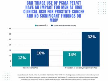
Study: PSMA PET/CT Reduces Biopsy Rate by Nearly 50 Percent for Men with Equivocal or Non-Suspicious Prostate mpMRI
5