
|Articles|February 26, 2007
Philips Medical rolls out advanced tools at HIMSS 2007
Philips’ service model of PACS got a shot in the arm at the Healthcare Information and Management Systems Society conference with the unveiling today of advanced visualization tools that will bring 3D to any desktop on the client’s iSite network. The tools, presented as works-in-progress due for general release later this year, will provide multiplanar reformats, maximum intensity projection, and other volume visualization capabilities.
Advertisement
Philips' service model of PACS got a shot in the arm at the Healthcare Information and Management Systems Society conference with the unveiling today of advanced visualization tools that will bring 3D to any desktop on the client's iSite network. The tools, presented as works-in-progress due for general release later this year, will provide multiplanar reformats, maximum intensity projection, and other volume visualization capabilities.
With its acquisition two years ago of Stentor, Philips Medical Systems chose to enter the PACS arena using a service model. Stentor's rebranded and evolved iSite Enterprise operates for Philips clients on a pay-per-study basis. Its core technology, the iSyntax, enables on-demand, enterprise-wide delivery of images to standard PCs and workstations using existing information technology infrastructure.
What's been lacking has been access to 3D images through these desktop PCs. Philips' answer, which debuted today at HIMSS, is now being evaluated at clinical sites, according to Sybo Dijkstra, senior director of marketing for Philips healthcare informatics.
It will be generally available by the end of 2007, he said. When it is, these tools will leverage the company's iSyntax, delivering volumetrically reconstructed images to desktops within three seconds. The speed comes from the ability of iSyntax to navigate large data sets to find and download only the needed data when they are needed.
"We have taken the platform radiology PACS in iSite Enterprise and integrated our clinical value so that you don't need a separate parallel infrastructure or special attachment to the PACS," Dijkstra told DI SCAN.
Gone is the additional thin-client server that some vendors require to do advanced postprocessing, he said. No need for a dedicated 3D workstation either.
"In this, 3D comes as an advanced functionality to virtually all users who want to have access to it throughout the healthcare enterprise," he said.
Users of these tools will also find they have access to more than just volumetric reconstruction. The MIP and MPR functions are enhanced with links between the 3D images and 2D representations. Dijkstra describes these links as "much more advanced than the standard MIP/MPR functionality present in most PACS," a claim he and his colleagues plan to demonstrate the rest of this week on the HIMSS exhibit floor.
Advertisement
Related Content
Advertisement


Advertisement
Advertisement
Trending on Diagnostic Imaging
1
SNMMI: What Early Research Reveals About the Alpha-Emitting Radioconjugate ATNM-400 for Prostate Cancer
2
Mammography Study Shows Impact of AI-Powered Slab Reconstruction with DBT
3
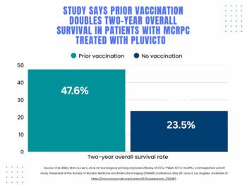
SNMMI: Does Vaccination Enhance Pluvicto Efficacy in mCRPC?
4
Molecular Imaging in Focus: PSMA PET Radiotracers and Urinary Radioactivity: What Head-to-Head Prospective Multicenter Research Reveals
5