Can Positron Emission Mammography Have an Impact in Diagnosing Invasive Breast Cancer?
Findings from a new pilot study showed that low-dose positron emission mammography (PEM) detected 96 percent of malignant index lesions and had a 46 percent lower false-positive rate in comparison to breast MRI.
Could low-dose positron emission mammography (PEM) provide a viable alternative in breast cancer detection?
In a prospective pilot trial, recently published in Radiology: Imaging Cancer, researchers assessed the use of low-dose PEM in 25 women (median age of 52) with newly diagnosed breast cancer. The study authors utilized different dosing of fluorine 18-labeled fluorodeoxyglucose (18F-FDG) with five patients dosed at 37 MBq, 10 patients dosed at 74 MBq and 10 patients dosed at 185 MBq, according to the study. All of the patients had concurrent magnetic resonance imaging (MRI) as well.
The researchers found that low-dose PEM diagnosed 24 out of 25 malignant index lesions, including 19 invasive cancers and five cases of in situ disease. Overall, PEM demonstrated an 87.1 percent sensitivity rate, a 94.7 percent specificity rate and a 90 percent accuracy rate, according to the study.


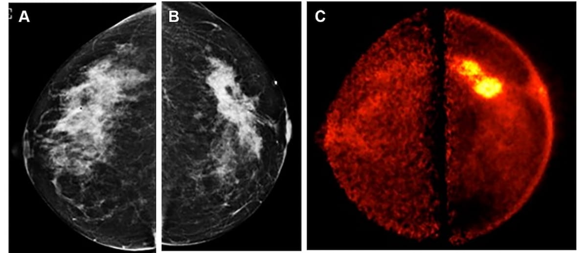
Here one can see mammography images (A and B) and a positron emission mammographic (PEM) view (C) for a 50-year female with a grade 2 invasive lobular carcinoma in the left breast. Note the 7.0 cm spiculated, irregular mass (B) and the intense uptake of fluorine 18-labeled fluorodeoxyglucose (18F-FDG) in the left breast on the PEM image. (Images courtesy of Radiology: Imaging Cancer.)
The researchers also noted the consistent detection of malignancies with PEM at the different dosing levels of 185 MBq and 74 MBq with the only missed diagnosis (a 38 mm lobular index cancer) occurring with the 37 MBq dosing.
“Although we did not observe significant differences among the various low FDG doses, most likely due to the limited sample size underpowering the study, it is noteworthy that we successfully reduced the dose to 37–185 MBq with PEM. This reduction corresponded to a decrease in radiation exposure of 0.62–0.71 mSv to 1.24–1.42 mSv. Notably, the decrement in dose did not hinder any detection of invasive disease, suggesting the plausible feasibility of utilizing lower FDG doses with this PEM device without compromising effectiveness,” wrote lead study author Vivianne Freitas, M.D., who is affiliated with the Joint Department of Medical Imaging with the University Health Network and the Sinai Health System at the Women’s College Hospital at the University of Toronto in Canada, and colleagues.
While breast MRI identified all 25 malignant index lesions, the study authors found that with additional lesion detection, PEM had a lower false positive rate (16 percent) in comparison to breast MRI (62 percent). Acknowledging the need for larger prospective trials to further explore and validate these findings, the researchers noted the potential of PEM for reducing extraneous follow-up imaging.
“These results may represent a potential added benefit of PEM, possibly decreasing both patient anxiety and the cost burden of the health care system related to additional imaging workups, including short-interval follow-ups and/or biopsy,” maintained Dr. Freitas.
(Editor’s note: For related content, see “Is Contrast-Enhanced Mammography a Viable Option for Diagnosing Invasive Lobular Carcinoma?,” “Mammography Surveillance: Can Screening DBT Have an Impact After Breast Cancer Treatment?” and “Study: Abbreviated MRI and DBT Offer Comparable Breast Cancer Detection in Dense Breasts.”)
In regard to study limitations, the researchers acknowledged that a small sample size, subjective visual parameters for lesion assessment and limiting the cohort to those who had a preoperative MRI may hamper extrapolation of these findings to a broader patient population. The study authors also suggested that awareness by radiologist readers that study participants had at least one cancer may have factored into the results.
Newsletter
Stay at the forefront of radiology with the Diagnostic Imaging newsletter, delivering the latest news, clinical insights, and imaging advancements for today’s radiologists.


The Reading Room Podcast: Current Perspectives on the Updated Appropriate Use Criteria for Brain PET
March 18th 2025In a new podcast, Satoshi Minoshima, M.D., Ph.D., and James Williams, Ph.D., share their insights on the recently updated appropriate use criteria for amyloid PET and tau PET in patients with mild cognitive impairment.

New PET Study Links Higher Education Level to Speed of Tau Accumulation with Alzheimer’s Disease
July 8th 2025For patients with amyloid β (Aβ)-positive findings on positron emission tomography, higher educational attainment was associated with accelerated accumulation and spread of tau, according to new research.


Considering Breast- and Lesion-Level Assessments with Mammography AI: What New Research Reveals
June 27th 2025While there was a decline of AUC for mammography AI software from breast-level assessments to lesion-level evaluation, the authors of a new study, involving 1,200 women, found that AI offered over a seven percent higher AUC for lesion-level interpretation in comparison to unassisted expert readers.


