
|Articles|June 14, 2006
‘Rise’ in thyroid cancers comes from better detection methods
More sophisticated detection methods account for a dramatic increase in thyroid cancer over the last 30 years, rather than a true change in occurrence of the disease, according to a study in the Journal of the American Medical Association. Physician self-referral may also be a contributing factor.
Advertisement
More sophisticated detection methods account for a dramatic increase in thyroid cancer over the last 30 years, rather than a true change in occurrence of the disease, according to a study in the Journal of the American Medical Association. Physician self-referral may also be a contributing factor.
Radiation is a common cause of thyroid cancer, and some have questioned whether the increase in thyroid cancer incidence was linked to greater exposure to radiation in the environment.
But stable mortality rates despite the apparent rise in incidence led researchers to conclude that the increase is actually due to better detection, not greater occurrence of disease.
Dr. Louise Davies and Dr. H. Gilbert Welch of the VA Outcomes Group and Dartmouth Medical Center investigated causes behind the increase of thyroid cancer incidence from 3.6 per 100,000 in 1973 to 8.7 per 100,000 in 2002 (JAMA 2006;295:2164-2167).
The 2.4-fold increase is mostly due to the detection of subclinical papillary cancers 2 cm or smaller. During the 30-year span, the incidence of papillary cancers grew from 2.7 to 7.7 per 100 000, a 2.9-fold increase.
The researchers found no significant change in incidence of the less common histological types: follicular, medullary, and anaplastic. In 2002, for example, papillary cancers accounted for 88% of all thyroid cancers, while 9% were follicular, and 3% were either medullary or anaplastic.
"Given the known prevalence of small asymptomatic papillary thyroid cancers at autopsy, we believe this suggests that increased diagnostic scrutiny has caused an apparent increase in incidence of cancer rather than a real increase," the authors said.
The use of ultrasound for thyroid disease became widespread in the 1980s, while fine-needle aspiration flourished in the 1990s. Ultrasound can detect nodules as small as 0.2 cm. FNA, which can be performed quickly during an office visit, replaced lengthy nuclear medicine scans.
"The ability to detect small nodules and then aspirate their contents has clearly facilitated the diagnosis of these smaller cancers," the authors said.
In fact, overdiagnosis of the disease has itself become a concern. While many people may die with papillary nodules, they do not die from them. Yet 75% of those with papillary cancers less than 1 cm undergo total thyroidectomy. This procedure carries small but significant risks of complications and requires lifetime maintenance and surveillance.
This concern is growing as thyroid ultrasound moves from the radiology department to physicians' offices. When ultrasound is used as an office-based adjunct to physical examination, the likelihood of self-referral increases, along with the number of nodules - and, ultimately, cancers - found and treated.
The authors suggest that follow-up for symptomatic thyroid nodules may be an appropriate course and that papillary cancers smaller than 1 cm could be classified as a normal finding.
They conclude that further research is needed to determine appropriate management of small papillary cancers detected with ultrasound and fine-needle aspiration. Data for the study came from Surveillance Epidemiology and End Results (SEER) and the National Vital Statistics System.
For more information from the Diagnostic Imaging archives:
Ultrasound helps reveal vascular patterns in thyroid cancer
http://www.dimag.com/showArticle.jhtml?articleID=170704231
Advertisement
Related Content
Advertisement


Advertisement
Advertisement
Trending on Diagnostic Imaging
1
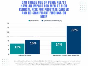
Study: PSMA PET/CT Reduces Biopsy Rate by Nearly 50 Percent for Men with Equivocal or Non-Suspicious Prostate mpMRI
2
Addressing Challenges in Radiology Reporting
3
Mammography Study: Can AI Detect Potential Breast Cancer Up to a Decade Prior to Diagnosis?
4
Diagnostic Imaging's Weekly Scan: June 7 — June 13
5