
|Articles|December 1, 2002
Software tool allows radiologists to flesh out electronic medical record
The development of a software tool using extensible markup language (XML) has allowed researchers in radiology informatics at the University of Pittsburgh Medical Center to add visible light images to their PACS archive. But the availability of a digital
Advertisement
The development of a software tool using extensible markup language (XML) has allowed researchers in radiology informatics at the University of Pittsburgh Medical Center to add visible light images to their PACS archive. But the availability of a digital mode of storing and transferring such images may pose security problems.
Because the DICOM standard supports visible light image files such as jpeg and tiff files, radiologists can add anatomic photographs into their electronic medical record to view alongside standard radiology images.
SimpleDICOMWrapper software developed by Steven Uttecht, a researcher at UPMC, enables radiologists to input visible light images along with pertinent metadata such as patient information and time of examination and to integrate that information into an archive.
"With DICOM, a $200 digital camera becomes a digital modality," he said.
The increasing ease of using digital media, such as digital cameras, makes it tempting to capture images anywhere, anytime. The pitfalls to such ease and ubiquity of image capture lie in managing and organizing the data.
"We've leveraged our preexisting PACS for storage and management of digital photos, which is extremely advantageous," Uttecht said.
The software provides an XML-derived interface, which allows users to organize jpeg and tiff files alongside the relevant metadata. Uttecht and colleagues initially demonstrated their system on dermatology photos created for skin cancer patients.
Using the software, the researchers were able to input visible light images, such as full-body photos of a selected patient, and arrange them alongside such metadata as anatomical maps, patient identification information, and previous patient history.
It is extremely important to take care when entering the metadata, as there is no database against which that metadata is checked, acording to Uttecht.
"We need to trust our metadata," he said. "The key is efficiency. How can we use the power of XML to not only organize metadata, but to ensure the integrity of that data?"
One of the biggest issues that crops up with the input of visible light images into a PACS archive is the question of security and privacy. Radiological images like CT and ultrasound scans are not immediately identifiable with a specific person, Uttecht said. With the digitization of visible light images, full-body photos as well as photos of a person's face can now be input and accessed from a PACS.
Uttecht acknowledged these issues during the question and answer session of his presentation and said that the researchers were looking into security measures such as limiting access to the data to only certain physicians and IP addresses.
Ultimately, the goal of such software implementation and visible light image integration is to round out the information available electronically, enabling physicians to view visible light images alongside radiology images.
"The goal is not just to store images but to complete the electronic health record," Uttecht said.
Advertisement
Related Content
Advertisement


Advertisement
Advertisement
Trending on Diagnostic Imaging
1
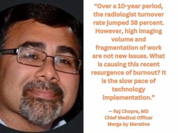
Five Strategies to Facilitate Technology Implementation and Alleviate Radiologist Turnover
2
Molecular Imaging in Focus: Key Takeaways from the 2026 SNMMI Conference with Michael Hofman, MBBS, FRACP
3
FDA Approves Low-Dose MRI Contrast Agent Gadoquatrane
4
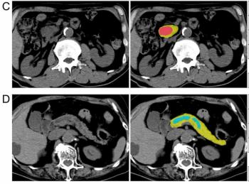
Can AI Enhance Non-Contrast and Contrast-Enhanced CT Detection of Pancreatic Cancer?
5