Studies test new breast tomosynthesis prototypes
News that two views are better than one with the emerging and promising technology of breast tomosynthesis raises questions about the technique’s practicality and cost-effectiveness as a screening tool.
News that two views are better than one with the emerging and promising technology of breast tomosynthesis raises questions about the technique's practicality and cost-effectiveness as a screening tool.
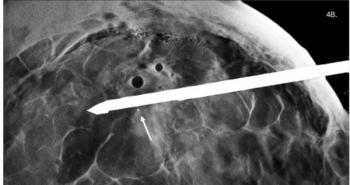
Tomosynthesis is a 3D digital mammographic technique that has been in development since the 1960s. In breast imaging, it minimizes the effects of overlapping tissue, possibly helping to improve sensitivity and specificity relative to conventional mammography.
Researchers at Massachusetts General Hospital, the technique's main birthplace, released a new study at the RSNA meeting stressing the importance of imaging the breast in both the craniocaudal and mediolateral oblique positions when performing tomosynthesis.
Some tomosynthesis devices involve 10 to 15 separate image acquisitions along an arc and one model acquires continuously along the arc. The technology has been touted as a technique that acquires information from one view (direction).
It now appears that two separate views, or two arcs, are needed. The tube is positioned lateral to medial and then moved to the cranial caudal direction. This would double the acquisition time and number of images, as well as make interpretation more time-consuming and require a higher dose.
Performing two views instead of just one would have a significant impact on workload and workflow, but the technique could potentially eliminate the need for other diagnostic exams and find cancers at an earlier stage.
"The need for two projections does complicate the situation. We will have to look carefully at costs and benefits," said Dr. Daniel Kopans, a professor of radiology at MGH, during a tomosynthesis session on Tuesday.
Although mammography has had a major positive impact on women's health in terms of reducing breast cancer mortality rates, it has much room for improvement with respect to sensitivity and falters in imaging dense breasts. Tomosynthesis has emerged as a glamorous digital contender for a screening role in the future, and relegation to a supporting spot in diagnosis would disappoint researchers in the field.
The MGH study involved 34 women scheduled to undergo biopsy. MLO and CC views were obtained with a Hologic tomosynthesis system at a total dose roughly equivalent to a standard mammogram. Researchers compared visibility of lesions on the different acquisitions.
Of 34 lesions detected, 16 were masses, three were masses with calcifications, seven were purely calcifications, and eight were cases of architectural distortion. Malignant histology was found in 68% of cases.
Both views enabled equally good visualization of 22 lesions. Four lesions were much more visible on the MLO view, however, and five were much more visible on the CC view. Three lesions were visible only on the CC view, and all of these proved to be malignant.
"When performing tomosynthesis, it is desirable to get both the MLO and CC views to optimize visualization of lesions," said Dr. Elizabeth Rafferty, an instructor in radiology at MGH, who presented the results.
The RSNA session also featured positive trial results with breast tomosynthesis for the detection and characterization of masses.
Dr. Joseph Lo, an assistant professor of radiology at Duke University Medical Center, presented promising research with one of the first Siemens Medical Solutions tomosynthesis prototypes. To date, Duke has imaged 184 patients, and Lo shared results for the first 144.
Researchers assessed sensitivity for lesion detection in 271 breasts. There were 30 lesions, including eight cancers. Mammography picked up 21 of 30 lesions, equivalent to a 70% sensitivity rate, whereas tomosynthesis picked up 27 lesions, or 90% sensitivity.
Tomosynthesis achieved this high sensitivity with a lower callback rate relative to conventional mammography (10% versus 15%), which indicates possible benefits for elimination of unnecessary biopsies and further workup.
Researchers at the University of Michigan Hospitals also reported positive results with tomosynthesis in imaging masses, using GE Healthcare's combined whole-breast ultrasound and tomosynthesis prototype system, which allows ultrasonic and mammographic imaging in a single acquisition.
The study involved 30 patients with nonfatty breasts. Two views were taken. Lesion size ranged from 4 to 50 mm, with a median size of 12 mm. Aside from one microcalcification case, all were masses. Mean patient age was 47 years, and there was a high percentage of dense breasts (68%). Dedicated, experienced breast radiologists performed the studies, blinded to final outcomes.
Of 24 lesions, tomosynthesis picked up 23, whereas only 20 were picked up with conventional mammography.
"Tomosynthesis detected more masses than conventional mammography and overall was preferred by the breast imagers," said Dr. Mark Helvie, a professor of radiology at Michigan, who presented the results.
Although these trials of the budding technology show positive results, breast imaging expert Dr. Carl D'Orsi, moderator of the session, noted that the studies involved mainly masses and said further work is needed on the more challenging use of tomosynthesis in calcifications. During a panel discussion, D'Orsi, chief of breast imaging at Emory University, questioned whether the technique would work as well in calcifications.
In response, Kopans said that calcifications are visualized as well if not better with tomosynthesis, but improvement in reader perception is needed to pick up calcification clusters. New techniques have been developed to aid in the detection of calcifications, and computer-aided detection could also play an important role in this respect, panelists said.
Newsletter
Stay at the forefront of radiology with the Diagnostic Imaging newsletter, delivering the latest news, clinical insights, and imaging advancements for today’s radiologists.


