Emerging research shows that photon-counting detector computed tomography (PCD-CT) led to shorter image acquisition time, significantly reduced radiation dosing and a more than 60 percent higher percentage of artifact-free examinations in comparison to energy-integrating detector CT (EID-CT) for the workup of suspected pulmonary embolism (PE).
For the retrospective study, recently published in European Radiology, researchers compared the use of EID-CT (158 patients in group 1) and PCD-CT (172 patients in group 2) in assessing suspected acute pulmonary embolism. The EID-CT cohort was comprised of two subgroups with group 1a (105 patients) receiving dual-energy EID-CT and group 1b (53 severely dyspneic patients) having single-energy EID-CT, according to the study.
In comparison to patients who had dual-energy EID-CT, the researchers found that PCD-CT led to lower mean image acquisition time (0.9 seconds vs. four seconds) as well as significant reductions in CT dose index volume (CTDIvol) (5.1 mGy vs. 9.5 mGy) and dose-length product (DLP) (169 mGy-cm vs. 323.4 mGy-cm).
“These results are in line with those recently reported in oncologic populations in which the radiation dose of PCD-CT examinations was significantly reduced compared to examinations acquired with comparable protocol settings on second-generation dual source CT equipment. In our study, the CTDIvol and DLP in (the PCD-CT group) were reduced by 46.3% and 47.7%, respectively, compared to (the dual-energy RID-CT group),” wrote lead study author Martine Remy-Jardin, M.D., Ph.D., who is affiliated with the Department of Thoracic Imaging at the University of Lille in Lille, France, and colleagues.
While mean image noise was higher in the PCD-CT cohort in comparison to patients who had dual-energy EID-CT (20.3 HU vs. 17.3 HU), the researchers found that the percentage of artifact-free exams with PCD-CT was over 60 percent higher than that of dual-energy EID-CT (89 percent vs. 28.6 percent).
Three Key Takeaways
- Reduced radiation dosing and image acquisition time. Photon-counting detector computed tomography (PCD-CT) offers substantial benefits over energy-integrating detector CT (EID-CT) in suspected pulmonary embolism (PE) cases. PCD-CT demonstrated significantly shorter image acquisition times (0.9 seconds vs. four seconds) and notable reductions in both CT dose index volume (CTDIvol) and dose-length product (DLP), leading to a decrease in radiation exposure by approximately 46-48 percent compared to dual-energy EID-CT.
- Higher percentage of artifact-free examinations. PCD-CT exhibited a significant improvement in image quality, with over a 60 percent higher percentage of artifact-free examinations compared to dual-energy EID-CT. Motion artifacts, particularly cardiogenic motion artifacts, were substantially reduced in PCD-CT scans due to the faster data acquisition speed, resulting in nearly 90 percent of PCD-CT examinations being free of perceptible motion around the left ventricle.
- Advantages of low-energy images. Utilizing lower-energy images in PCD-CT can overcome limitations related to transient interruption of contrast and/or a patent foramen ovale, resulting in improved diagnostic accuracy. Retrospective reading of low-energy images in all patients scanned with PCD-CT provides a significant advantage in daily routine, previously unavailable in a considerable portion of patients scanned with EID-CT.
“Regarding motion artifacts, the speed of data acquisition in (the PCD-CT group) explains the significant difference in the distribution of cardiogenic motion artifacts between (the PCD-CT group) and (the dual-energy EID-CT group) with no perceptible motion around the left ventricle in 89.0% of (PCD-CT) examinations, a result very close to the 90.6% of (dual-energy EID-CT) examinations free of cardiogenic artifacts,” pointed out Remy-Jardin and colleagues.
(Editor’s note: For related content, see “Is Photon Counting CT a Better Option than Dual-Energy CT for Pulmonary Angiography?,” “Study Says Photon-Counting CT Offers Better Lung Assessment than Conventional CT” and “Study: Photon-Counting CT for Pulmonary Angiography Allows Significant ICM Reduction.”)
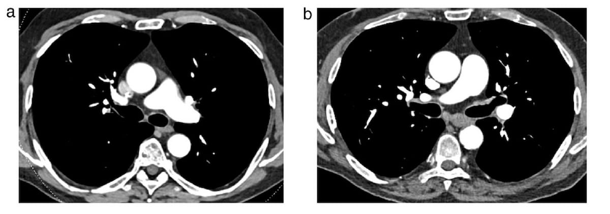
When there is opacification of pulmonary arteries caused by transient interruption of contrast and/or a patent foramen ovale, the study authors suggested that utilizing lower-energy images can overcome this limitation.
“The retrospective reading of low-energy images in all patients scanned with PCD-CT is an obvious advantage in daily routine, previously inaccessible in more than one-third of patients scanned with EID-CT,” added Remy-Jardin and colleagues.