
|Articles|October 3, 2012
Subspecialist Readings of Pediatric Images Improves Care
Author(s)Marijke Vroomen Durning, RN
Imaging study interpretations by subspecialty radiologists provide important clinical information about pediatric patients.
Advertisement
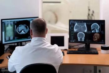
Imaging study interpretations by subspecialty radiologists provide important clinical information about pediatric patients and should be recognized as integral to optimal care, according to a study in the October issue of American Journal of Roentgenology.
Researchers from Gunderson Lutheran Medical Center in La Crosse, Wis., sought to identify if there were differences in imaging interpretations between radiologists from referring centers and radiologists at a tertiary care children’s hospital.
A total of 773 examinations performed on children between January 1, 2009, and May 31, 2010, were retrospectively reviewed for comparisons between the original and second interpretations. The cases were each categorized by a fellowship-trained pediatric radiologist and a neuroradiologist. They used the content of the two radiology reports as agreement versus minor or major disagreement.
Researchers found that the tertiary care radiologists provided more accurate readings than did radiologists from outside the institution.
There were disagreements between interpretations in 323 of the 773 reports (41.8 percent), of which 168 (21.7 percent) were major and 155 (20 percent) were minor. Neurologic studies were most frequently requested for reinterpretation, 427 (55.2 percent), most commonly in the setting of trauma, 286 (67.0 percent). Major and minor disagreements about the 427 neuroimaging cases occurred in 54 (12.6 percent) and 91 (21.3 percent), respectively.
“Our findings suggest that discrepancy rates for second interpretations in studies of pediatric patients transferred to tertiary care pediatric institutions are substantial,” concluded the authors. “These results indicate that interpretations by subspecialty radiologists at a point-of-care facility provide important clinical information about the pediatric patient and should be recognized by payers as integral to optimal care.”
The major disagreements most frequently observed concerned the presence of fractures and hemorrhages. Among 305 body imaging cases, major and minor disagreements occurred in 99 (32.6 percent) and 57 (18.7 percent) cases, respectively.
Concern for appendicitis was the most common setting for nontraumatic body imaging (168/305, or 55.1 percent) and the indication for imaging was responsible for 40.3 percent of major disagreements in nontraumatic abdominal imaging.
Requests for reinterpretation were rare for radiographic studies (5.3 percent), which had major and minor disagreement rates of 36.6 percent and 17.1 percent, respectively.
The majority of cases, 90.2 percent, showed the second interpretation was more accurate than the original.
Advertisement
Related Content


Advertisement
Advertisement
Trending on Diagnostic Imaging
1
FDA Approves Low-Dose MRI Contrast Agent Gadoquatrane
2
The Hidden Social Price of Remote Work in Radiology
3
Study: PSMA PET/CT Reduces Biopsy Rate by Nearly 50 Percent for Men with Equivocal or Non-Suspicious Prostate mpMRI
4
Addressing Challenges in Radiology Reporting
5