
|Articles|March 13, 2007
Wall-hung flat panels transform earthbound images at European Congress
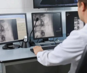
Philips and BrainLab unveiled their own brands of magic at the European Congress of Radiology, one creating an illusion of 3D, the other an interface relying on sleight of hand.
Advertisement
Philips and BrainLab unveiled their own brands of magic at the European Congress of Radiology, one creating an illusion of 3D, the other an interface relying on sleight of hand.
Building on technologies developed initially for consumers, Philips Medical Systems offered up a big-screen LCD with algorithms that turned flat images into 3D reconstructions that appeared to hang in midair.
BrainLab played its tricks using a similar wall-hung rectangular monitor that also featured a built-in touchscreen. Windows opened on screen displayed images that responded to the fingertips of an operator. Users could resize them by drawing two fingers together or apart, pan or move them around with a single finger, or scroll through a stack of slices, changing contrast or measuring from one point to another.
The Philips prototype, a passive display, allowed no such interactions but made up for it in wow. Flashing on screen, one after the other, were brilliant images of the brain with vasculature protruding, cutaways of the human skull, an aneurysm bulging from between two ineffective stents, and a volumetric reconstruction of the abdomen with an interventional tool thrust in it.
Impressive as each was, in the end, both displays were as much about seeking guidance for future direction as demonstrating engineering prowess. Separately, the two companies were seeking feedback from radiologists about how their technologies might be used. Sophie Perceval, Philips marketing manager for cardiovascular x-ray, noted that their technology is being examined for its value as a review medium rather than for diagnostic applications. The images take shape only when observed from a meter or more away, a distance necessary for the eye to appreciate the illusion of 3D. Images prepared in this way might provide physicians another perspective on data, but their true clinical value, if any, remains to be determined.
BrainLab strategists had not yet settled on whether the purpose of their Digital LightBox would be to review images, plan interventions, or even assist in diagnoses. Ultimately, the Digital LightBox might be tied into a hospital information network to call up virtually any patient information and display it anywhere. Its presence at the ECR was primarily to stimulate discussion, according to Christopher Hamilton, BrainLab project manager.
"We are developing a really intuitive and new way of interacting with images," he said. "We are trying to go to the core to make interaction as easy as possible."
Advertisement
Related Content
Advertisement


Advertisement
Advertisement
Trending on Diagnostic Imaging
1
What a Meta-Analysis Reveals About FAPI PET/CT for Detection of Peritoneal Metastases
2
FDA Clears CT-Based AI Software for Detection and Triage of Vascular Occlusion in Lower Extremities
3
Emerging PET Radiotracer May Enhance Detection of Small Metastases in Patients with Advanced Melanoma
4
Put it on Autopilot: Your Minimal Effort Guide to Radiological Greatness
5