
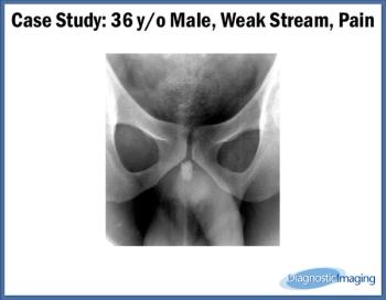
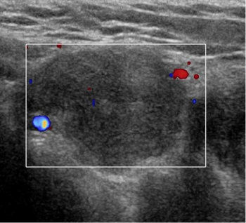
Case History: 36-year-old male presented with weak stream and pain.
Case History: 36-year-old male presented with weak stream and pain.
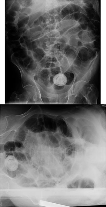
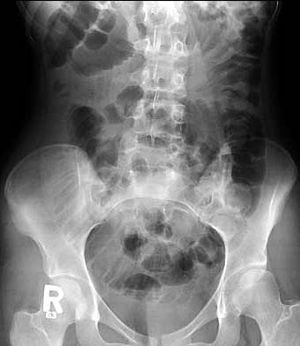
Incarcerated hernia: X-ray and CT scans evaluate inquinal swelling and and long-term swelling in elderly males.
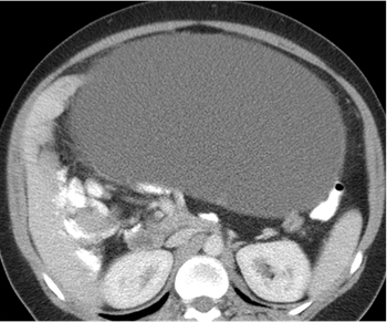
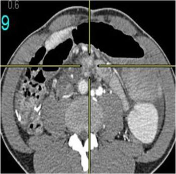
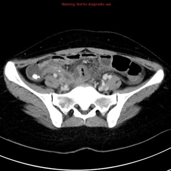
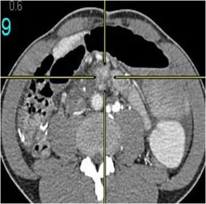
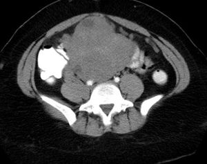
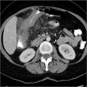
Mucinous cystadenom: 33-year-old female with abdominal distension and suspected ovarian mass. CT scan of abdomen and pelvis done with oral and intravenous contrast with multiple axial sections along with coronal and sagittal reformation.
A 45-year-old man with abdominal pain receives a CT angiogram. CT angiogram demonstrates acute angulation and narrowing of the proximal celiac axis.
A 73-year-old man with known case of liver cirrhosis receives a CT scan. A triphasic contrast study of the liver is performed.
A 69-year-old male patient presented with a three-week history of right upper quadrant abdominal pain, pyrexia.
Three cases: A 98-year-old male with multiple co-morbidities presented with abdominal pain, fever and distension; Elderly female patient with right upper quadrant pain; A 50-year-old female with acute abdominal pain, r/o obstruction.
An 11-year-old boy with frequent partial seizures and lip twisting for four years. Initial CT of brain was done without contrast as patient presented to ER. The patient needs further evaluation by MRI to rule out focal mass.
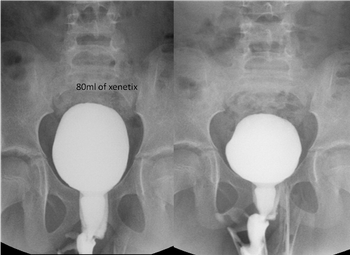
A 7-year-old girl with day and night urge incontinence. Voiding cystourethrogram performed. Low capacity of bladder and patient started voiding after 80ml of contrast injection in urinary bladder through catheter. Unusual dilatation of urethra, giving “spinning top” appearance.
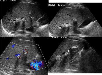
A 23-year-old male with alcohol consumption with elevated liver enzymes. Ultrasound shows intrahepatic ductal dilatation involving Seg 6 RLL with muliple echogenicity with posterior shadowing represent calculi.
A 12-year-old girl with bilateral wrist deformity, characterized by growth disturbance in the volar ulnar distal radial physis resulting in volar and ulnar tilted distal radial articular surface, volar translation of the hand and wrist and dorsally prominent distal ulna.
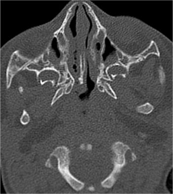
CT scan diagnoses right choanal atresia in 6-month-old male child. Pre-surgical evaluation. Non-enhanced axial CT scan of paranasal sinuses.
A 7-week-old female presents with left scalp hard swelling. The skull radiograph demonstrates a rim of calcification in the periphery of a raised swelling on the left parietal bone.
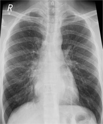
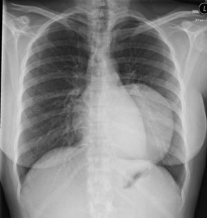
Clinical History: A 31-year-old man with back pain. X-Ray Chest: Lower thoracic paravertebral soft issue with trabeculated T9. Lungs are clear. Heart size within normal limit. No other obvious bony abnormality.
Clinical History: An 84-year-old male patient, known case of HTN and dyslipidemia, complaining of dysphasia for three months. Anorexia and vomiting. Diagnosed as esophageal adenocarcinoma outside for evaluation.
Clinical History: A 60-year-old female with pelvic fracture developed shortness of breath with tachycardia and tachypenea
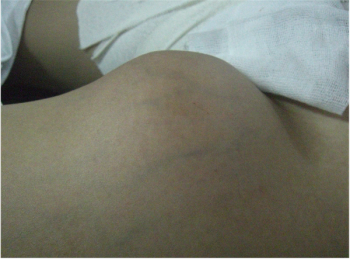
A 17-year-old male patient, no past medical history, presented with two-month history of left groin swelling with gradual increase in size and now pain.
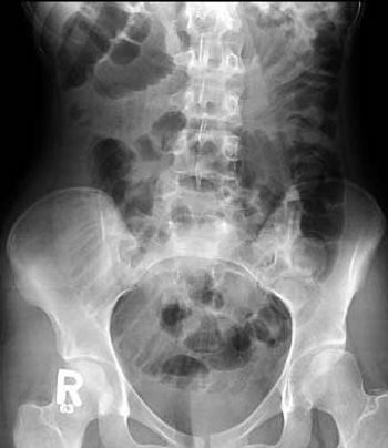
A 16-year-old female patient with repeated vomiting. First X-ray of the abdomen shows multiple dilated small bowel loops, represent distal small bowel obstruction. CT of the abdomen done for further evaluation.

A 56-year-old female presented with abdominal pain, R/O colitis. There is a long segment of asymmetrical wall thickening seen involving the ascending colon just above the ileocaecal region to proximal third of transverse colon associated with luminal narrowing and significant surrounding fat stranding and enlarged regional lymph node.
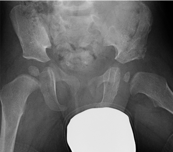
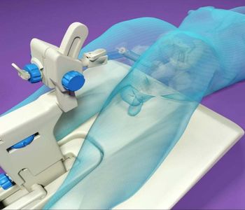
Developmental dysplasia of the hip (DDH) is a dislocation of the hip joint that is present at birth. The condition is found in babies or young children. A hip that is truly dislocated in an infant should be detected at birth, but some cases are mild and symptoms may not develop until after birth, which is why multiple exams are recommended.
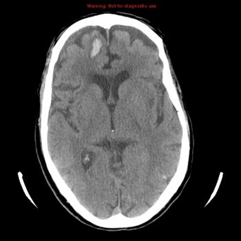
An 85-year-old male patient, medically free, complaining of a fall with trauma to right side of the head, and complaining of heavy tongue and right-sided weakness.
18-year old female presented in ER with complaint of right sided abdominal pain for two days associated with fever, vomiting, and diarrhea.
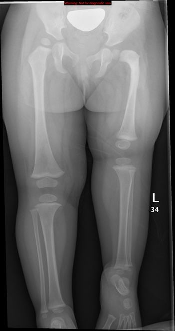
Newborn baby with short left lower limb with appearance of proximal femoral focal deficiency. Femur is shortened, flexed, abducted, and externally rotated.

Endometrial stromal sarcoma (ESS) is a rare malignant tumor of the endometrium, occurring in the age group of 40-50 years.

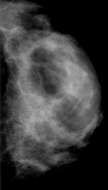
On routine chest screening X-ray, a 25-year-old, asymptomatic, female patient found to have well-defined, rounded soft tissue opacity in left lower thoracic para spinal region.
November 15th 2013
May 19th 2011
May 26th 2011
October 13th 2011
October 26th 2011

November 21st 2011