
|Articles|October 18, 2013
Ambient Light Affects Image Interpretation
Author(s)Diagnostic Imaging Staff
When interpreting images on handheld devices, ambient light should be a consideration for accurate readings.
Advertisement
The quality of images interpreted by radiologists using mobile devices may be affected by ambient light, according to a study published in the
The availability of apps to allow for image interpretation has given radiologists the ability to access images wherever they are. However, even if a particular device has been accepted as a reliable tool, its effectiveness may change according to ambient lighting levels. Researchers from the University of Maryland undertook and observational study to determine if these effects were significant.
The researchers asked three subjects to detect and distinguish four characters embedded in a white-noise background using two current-generation smartphones. The setup included five illumination conditions simulating dark room (super- and medium-dark), office (average), and outdoor (medium- and super-bright) environments.
The researchers found that the darker the lighting, the better the quality of the image interpretation.
“We found and quantified that due to the high reflectivity of handheld devices, performance deteriorates as the user moves from dark areas into environments of greater ambient illumination,” the authors wrote. “The quantitative analysis suggests that differences in display reflection coefficients do not affect the low illumination performance of the device but rather the performance at higher levels of illumination.”
Advertisement
Related Content
Advertisement


Advertisement
Advertisement
Trending on Diagnostic Imaging
1
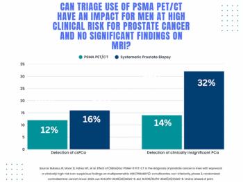
Study: PSMA PET/CT Reduces Biopsy Rate by Nearly 50 Percent for Men with Equivocal or Non-Suspicious Prostate mpMRI
2
Addressing Challenges in Radiology Reporting
3
The Hidden Social Price of Remote Work in Radiology
4
Mammography Study: Can AI Detect Potential Breast Cancer Up to a Decade Prior to Diagnosis?
5