
|Articles|January 1, 2001
Automated technique assures that PACS images match patients
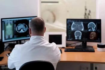
Maintaining the reliability of a PACS can be as simple as verifying that a patient's folder does not contain the wrong images. Without a way to do this automatically, verification of images becomes a time-consuming manual process. That may change,
Advertisement
Maintaining the reliability of a PACS can be as simple as verifying that a patient's folder does not contain the wrong images.
Without a way to do this automatically, verification of images becomes a time-consuming manual process. That may change, however, according to a paper presented at the 2000 RSNA meeting. Japanese researchers at the Kyoto Prefecture reported the development of an automated image-matching system that warns technicians when a wrong image may be present.
The study used an image database consisting of 2000 digital radiographs that included 1000 current and 1000 previous images for each of 1000 different patients. Results of preliminary testing of the image-matching system, designed to prevent the wrong chest radiographs from being stored in a patient's PACS file, indicate that approximately 50% of all wrong images can be correctly identified.
"We believe that this automated warning system for patient identification would be useful in correcting wrong images being stored in the PACS environment," said J. Morishita, Ph.D., a researcher at the Kyoto College of Medical Technology. "Physicians can now avoid reading the wrong patient's image during interpretation."
The strategy is to develop a correlation value for a previous image of the patient and a current image of the presumed corresponding patient, Morishita said. This is done by rotating the current image, shifting it horizontally and vertically to determine the best match between it and the previous image. This correlation value is then compared with a threshold value determined by analysis of histograms of correlation values obtained for the same patient and other patients.
If the correlation value is larger than the threshold, the current image will be considered to belong to the same patient and the image will be stored in the correct patient PACS folder, Morishita said. Conversely, if the correlation value is smaller than the threshold, the current image is considered to potentially belong to another patient and a warning is issued to alert radiology personnel to verify whether the current image really belongs to this patient.
Morishita said the correlation values between the current and previous images for the same "correct" patients were generally greater than those for "wrong" patients. Although the two histograms overlap at correlation values greater than 0.80, most parts of the histograms are separated. If the threshold value of 0.80 is selected for determination of wrong patients, the system provides 47.7% of true warning without any false warnings for the correct patient.
Advertisement
Related Content
Advertisement


Advertisement
Advertisement
Trending on Diagnostic Imaging
1
Molecular Imaging in Focus: Heather Jacene, MD, Charts Priorities as the New President of SNMMI
2
Can Breast Ultrasound AI Offer Comparable Sensitivity to Radiologists in Pregnant and Lactating Women?
3
FDA Approves Low-Dose MRI Contrast Agent Gadoquatrane
4
Addressing Challenges in Radiology Reporting
5