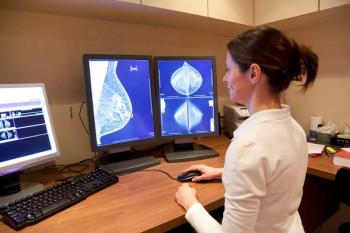
Breast CAD branches out from conventional mammography
Clinical acceptance of novel breast imaging techniques could hinge on concurrent development of appropriate computer-assisted detection packages, according to speakers at CARS.
Clinical acceptance of novel breast imaging techniques could hinge on concurrent development of appropriate computer-assisted detection packages, according to speakers at CARS.
Emerging approaches to breast cancer detection and diagnosis include digital breast tomosynthesis, contrast-enhanced MRI, and freehand ultrasound. Each of these techniques, however, involves a complex and lengthy image interpretation process.
In digital breast tomosynthesis, breast slices are reconstructed from a series of 11 projection images, said Dr. Robert Nishikawa, an associate professor of radiology at the University of Chicago. A single breast volume may contain about 50 slices, depending on breast thickness and slice spacing. So although the technique makes masses easier to see than on conventional mammograms, it also requires radiologists to interpret a much larger amount of data.
A collaborative project between the University of Chicago and Massachusetts General Hospital is addressing this issue. Researchers have developed a new preselection algorithm that provides accurate mass detection with reasonable efficiency. Tests on 36 tomographic breast volumes demonstrated 100% sensitivity with 23 false positives and 90% sensitivity with 13 false positives. The same team had developed an earlier system that attained just 81% sensitivity with 35 false positives.
The work represents an important first step toward development of CAD for digital breast tomosynthesis images, according to Nichikawa. Further refinements to eliminate false positives, based on feature analysis, should be developed as digital breast tomosynthesis itself evolves, he said.
"We need to evaluate our algorithm on a larger database, but that is hard to get. Right now, we think there are only a few working systems in the world, probably fewer than five. But manufacturers are starting to make more systems, so, hopefully, that will help provide a larger database," he said.
Speaking in the same session on breast CAD advances, Dr. Federica Pediconi, a radiology researcher at the University of Rome La Sapienza, showed results of a new DICOM-compatible software package designed to speed up tumor detection and reduce interoperator variability in the analysis of contrast-enhanced breast MRI. The semiautomated system takes just five minutes to complete its assessment of a breast exam.
The computerized tool first processes breast MRI data to obtain subtracted images. False color maps are then produced for each scan plane, based on known washout characteristics of benign, borderline, and malignant lesions.
Assessment of the software in 36 women with 68 histologically confirmed lesions (54 malignant) revealed excellent correlation with traditional methods of contrast-enhanced breast MRI evaluation. The two methods disagreed in just two cases, Pediconi said.
"Our software is visually intuitive, and it is also quite specific for lesions smaller than 10 mm in diameter," she said.
Freehand ultrasound is another promising tool for breast cancer diagnosis, according to Dr. Ruey-Feng Chang, a researcher in the department of computer science and information engineering at National Chung Cheng University in Taiwan. But a freehand scan of the whole breast, using a 2D ultrasound probe, can produce up to 2000 individual images for analysis.
Researchers from National Chung Cheng University, working with colleagues at Changhua Christian Hospital in Taiwan, Dokkyo University School of Medicine in Japan, and Seoul National University Hospital, have developed an automatic screening system to detect lesions from 2D breast ultrasound data.
The CAD system identified almost all tumors and cysts in 13 test cases. Additional trials are needed to verify its accuracy, and reduce false-positive calls, Chang said.
Newsletter
Stay at the forefront of radiology with the Diagnostic Imaging newsletter, delivering the latest news, clinical insights, and imaging advancements for today’s radiologists.


