
|Articles|October 20, 1993
California start-up firm targets mammography CAD
Computer-aided diagnosis of mammography exams was moved one stepcloser to commercial reality with the signing last month of alicensing agreement between the University of Chicago and R2 Technology.The California start-up company is raising financial
Advertisement
Computer-aided diagnosis of mammography exams was moved one stepcloser to commercial reality with the signing last month of alicensing agreement between the University of Chicago and R2 Technology.The California start-up company is raising financial backing andhopes to have a product in clinical trials within a year, accordingto Robert M. Foley, vice president of marketing.
Studies indicate that computer-aided diagnosis (CAD) can reduceby up to 50% the number of suspicious lesions missed by mammographersduring routine screening, according to Foley. R2 Technology's"second-reader" system is designed to reduce interpretationerrors by providing a backup to mammographers that never tiresor becomes distracted.
"The goal is to reduce the number of lesions that aremissed, to find breast cancer earlier," said Maryellen Giger,an associate professor of radiology and a member of the Universityof Chicago team that developed the system. "The radiologistis not perfect and neither is the computer, but combined theyshould have a better detection rate."
The system was developed at the Kurt Rossmann Laboratoriesfor Radiologic Image Research in the university's department ofradiology. In addition to Giger, other members of the team includelab director Kunio Doi, assistant professor Robert Nishikawa andDr. Robert Schmidt.
R2 Technology's system is based on IBM's RS/6000 workstation.The system also includes a film digitizer and backlit viewingstation.
Mammography films are first read by a radiologist, who makesa preliminary interpretation without help from the computer. Thefilms are then fed into the CAD system's digitizer, which sendsthe digitized data to the workstation for processing.
The CAD software detects possible abnormalities in the breast,such as microcalcifications or mass lesions, and alerts the mammographerby inserting an arrow on the workstation display screen that pointsto the suspicious area. The mammographer can then alter his orher interpretation of the mammogram depending on the suggestionsof the CAD program.
The workstation uses several different computer algorithmsto accomplish its task. To detect microcalcifications, the systemuses a spatial filtering method called a difference-image technique,Giger said. The microcalcifications are first enhanced to removevariations in background density due to normal structures. Afterfeature analysis, microcalcifications are indicated by an arrowon the workstation's CRT screen.
Mass lesions are detected using several techniques, includinga nonlinear subtraction processing method that compares the symmetryof the right breast to the left. The program also examines thegradient of the edges around the lesion to determine whether asuspicious mass is a real lesion or a natural asymmetry.
The program employs a form of artificial intelligence, calledartificial neural networks, to distinguish real lesions from falseones, Giger said. Data from known cases have been input into thesoftware, and the program uses this experience to make judgmentson which areas to highlight.
The CAD program has a sensitivity of about 85% for both microcalcificationsand mass lesions. The program registers fewer than two false positivesper image when identifying microcalcifications and fewer thanone false positive per image when identifying mass lesions, accordingto Giger.
Mammography CAD could come into its own when linked to digitalmammography screening units such as those under development byLorad and Fischer Imaging (see story page 3). CAD software couldbe installed on these units, allowing mammographers to see CADinterpretations on the unit's display screen simply by flippinga switch or pushing a button.
The University of Chicago team will display the CAD systemat the scientific exhibit at this year's Radiological Societyof North America meeting. This will be the fourth year the systemhas been shown at the conference, Giger said.
In addition to seeking financial backers, R2 Technology islooking for office space in the San Francisco Bay area, Foleysaid. Prior to R2 Technology, Foley worked for Integrated SurgicalSystems, a company developing a robotic surgery system using technologylicensed from IBM.
R2 Technology's president and CEO is S.P. Bob Wang, who developeda low-dosage rare-earth x-ray intensifier screen that was soldto 3M.
R2 Technology expects to price the CAD system at about $125,000,Foley said.
While some mammographers might not welcome the idea of a siliconchip looking over their shoulder, radiologists who have seen thetechnology at past RSNA meetings have been receptive to it, andGiger believes that CAD will be welcomed by most mammographers.
"You could think of this as another tool--in this caseit's in imaging--as opposed to analyzing some blood sample,"Giger said. "The final decision is made by the radiologist.They will appreciate extra pointers in (finding suspicious) areasthey might not have found."
BRIEFLY NOTED:
- Mobile MRI provider MTI's long journey down the restructuringroad is coming to an end. The Los Angeles company announced lastweek that it has reached an agreement in principle with lendersand stockholders that reduces debt and strengthens its balancesheet. The agreement will substantially reduce MTI's interestpayments and will include a debt-to-equity conversion, accordingto the company. The agreement was reached after negotiations betweenequipment suppliers, lenders and majority owners.
MTI operates the world's largest fleet of mobile MRI vans.The company was hit hard when demand for mobile services leveledout as many customers moved to acquire their own scanners. MTIhad been in discussions with lenders to renegotiate its debt sincelast year, and accelerated those talks in January (SCAN 4/7/93).
- Raytheon of Lexington, MA, has reentered the medical imagingfield through a joint telemedicine effort with Interactive TelemedicalSystems of Coral Gables, FL, and cardiac surgeon Michael E. DeBakeyof Texas Medical Center in Houston. Remote diagnosis will be apart of the MedTel interactive video system introduced by thethree parties last month.
Raytheon, a $9 billion electronics and defense conglomerate,withdrew as a supplier of medical imaging components and systemsfive years ago (SCAN 10/12/88). The firm's Machlett Laboratories,an independent x-ray tube manufacturer, was sold to competitorVarian. Raytheon's x-ray systems business was sold to FischerImaging and its distribution relationship with Hitachi in nuclearmedicine was passed on to Summit World Trade.
- ADAC Laboratories has agreed in principle to acquire radiologyinformation systems vendor SD&G Healthcare Systems. ADAC,of Milpitas, CA, will pay approximately $3.8 million for the SantaClara, CA, company. SD&G registered sales of $4 million lastyear.
SD&G will be combined with ADAC's information systems businessunit into a wholly owned subsidiary of ADAC. The new subsidiarywill be managed by Steven J. Dennis, president and CEO of SD&G.The new subsidiary will continue to market and support both theADAC MARS II and SD&G Images/3000 radiology information systemproducts.
- Monoclonal antibody developer Cytogen announced last weekthat president Thomas J. McKearn will take on the additional positionof CEO effective January 1994. Current CEO George W. Ebright willcontinue in an active management role as chairman long enoughto assure a smooth transition.
Slow sales of Cytogen's OncoScint monoclonal antibody agentforced the company to restructure last month (SCAN 10/6/93). Whilethe Princeton, NJ, company will continue to promote OncoScint,it will place new emphasis on developing peptide-based imagingagents.
- CTI Services opened its second PET compound distributioncenter (PCDC) last month at Columbia Presbyterian Medical Centerin New York City. PET radiopharmaceuticals will be manufacturedon site using a Siemens cyclotron and related equipment. The NewYork PCDC will offer radiopharmacy consulting services, as wellas maintaining a radiopharmacy utilizing a board-certified nuclearpharmacist.
CTI Services' first PCDC was established in Tampa. Two sitesin Dallas and Chicago are in the final stage of development.
Advertisement
Related Content
Advertisement


Advertisement
Advertisement
Trending on Diagnostic Imaging
1
Molecular Imaging in Focus: Key Takeaways from the 2026 SNMMI Conference with Michael Hofman, MBBS, FRACP
2
Can AI Enhance Non-Contrast and Contrast-Enhanced CT Detection of Pancreatic Cancer?
3
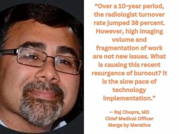
Five Strategies to Facilitate Technology Implementation and Alleviate Radiologist Turnover
4
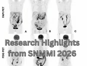
Research Highlights from SNMMI 2026
5