In the field of medical imaging, magnetic resonance imaging (MRI) stands out for its ability to provide high-resolution anatomical and functional images of internal body tissues. Despite its biological safety and non-invasiveness, the MRI procedure is often associated with symptoms of anxiety and discomfort in patients. Statistically, between 4 and 37 percent of patients experience anxiety during the examination with 1 to 15 percent exhibiting severe forms of anxiety, claustrophobia, or panic attacks. Moreover, 10 to 14.5 percent of MRI exams are prematurely terminated due to these symptoms.
Claustrophobia, a term derived from the Latin "claustrum" (closed space) and the Greek "phobos" (fear), is defined as an intense and irrational fear of enclosed and confined spaces. In the context of MRI, this phobia can present a significant challenge as the patient is confined inside the narrow bore of the scanner, an environment that can induce feelings of confinement and isolation.
The perception of anxiety by patients during MRI shows significant variations throughout the examination. Studies indicate that anxiety is highest at the beginning, tends to decrease during the scan, and may increase again toward the end. These variations can be influenced by various factors, including the duration of the examination, the noise generated by the scanner, and the patient's personal expectations. It is interesting to note that pre-examination anxiety can also be influenced by the patient's level of education with more educated individuals often experiencing higher levels of anxiety.
The first visual impression of an MRI scanner is a relevant variable associated with the occurrence of claustrophobia. Claustrophobic reactions are related to cognitions such as “harm caused by the machine.” Moreover, a considerable number of claustrophobic events, including premature termination of the MRI examination, occur upon merely looking at the scanner before the actual examination and experience of lying within the scanner have even started. Older generation scanners featuring closed, rather narrow, and long bores can also trigger a claustrophobic experience. Therefore, it has been suggested that patient comfort should drive the development of new scanners.
Patient information and education regarding the MRI procedure have been recognized as effective methods to reduce anxiety during the examination. To this end, new scanners (e.g., with wider and even shorter bores or with an open design that provide a lateral panoramic view) have been developed and are increasingly utilized. The use of such scanners in clinical routine contributed to the reduction of claustrophobic events and increased patient comfort in past studies.
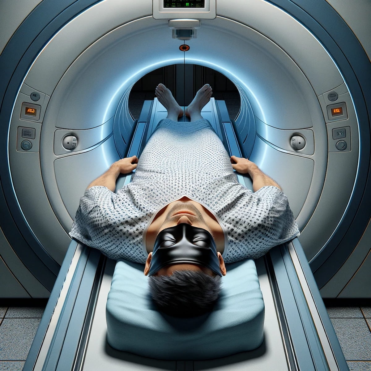
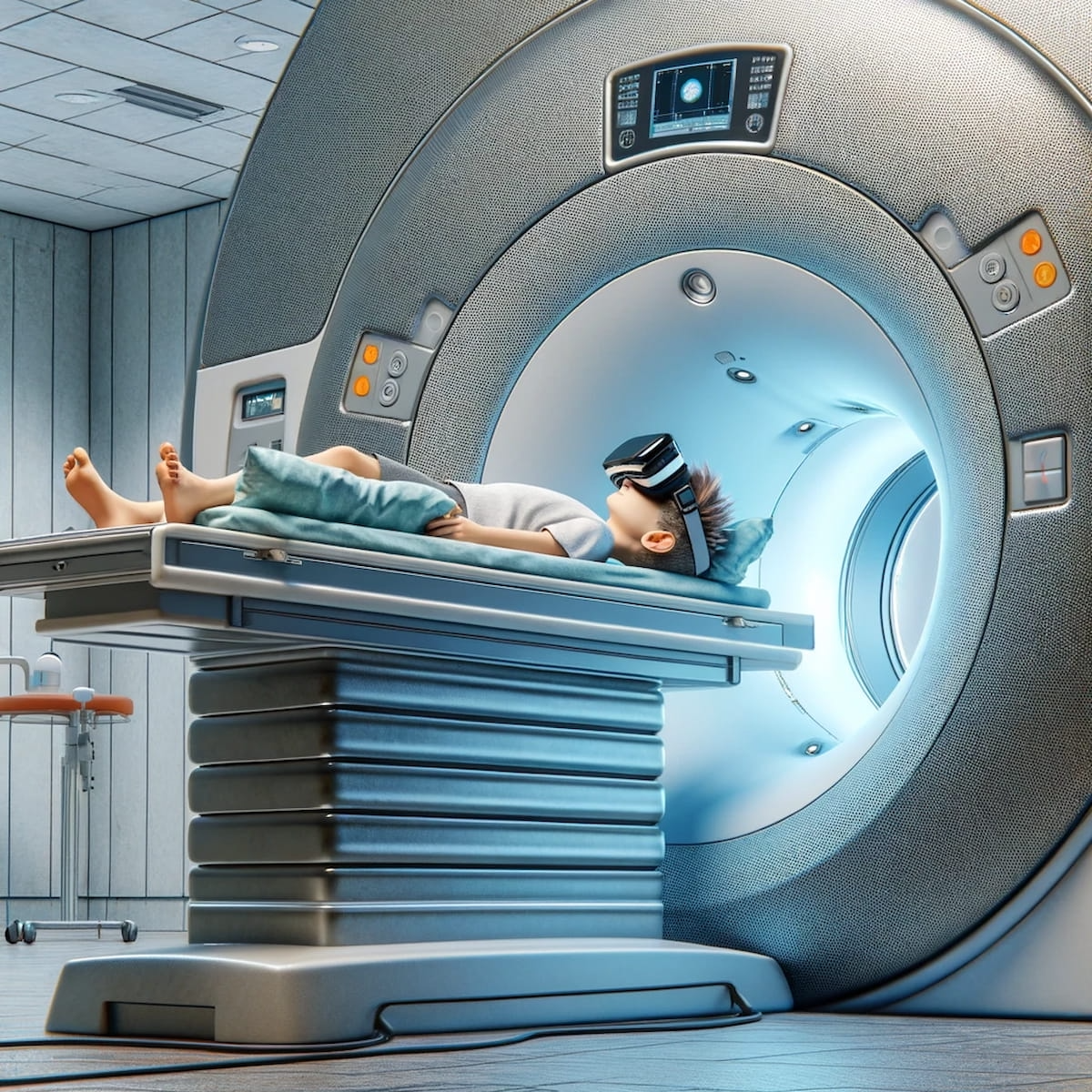
In recent decades, the field of MRI has witnessed significant technological innovations aimed at improving the patient experience, especially in managing claustrophobia. These advancements include developments in scanner design, radiofrequency coils, and the reduction of acoustic noise generated during scans. In order to help alleviate claustrophobia, many techniques such as the introduction of silent gradients, additional light in the bore, special glasses, and virtual reality tools have been successfully introduced into clinical practice.
Magnetic resonance imaging presents challenges including high noise levels and discomfort for patients. Noise, which can reach 130 dB, is particularly problematic for children, claustrophobic patients, and those with tinnitus or sensitive hearing. Reducing noise can help decrease exam-related anxiety and make the procedure more tolerable for patients. Accordingly, the use of earplugs or headphones for patients during MRI examination has become standard practice.
Emerging Solutions for Addressing Noise Reduction During MRI Exams
To further address the issue of noise in MRI scans, mechanical and sequencing solutions have been explored. Mechanical solutions include the use of damping materials around the gradient coils and placing the gradient coils in a vacuum to reduce acoustic noise.
Sequencing solutions involve adjusting and optimizing the movements of the gradient. However, reducing the response speed (slew rate) in conventional image acquisitions can lead to less efficient scanning and, in some cases, blurred images. To overcome these limitations, companies have developed alternative image acquisition techniques.
• Silent Scan (Silenz, GE HealthCare). This technique reduces acoustic noise by lowering the slew rate in comparison to conventional image sequences, producing noise of only 69 dB. Silent MRI uses a 3D gradient-echo imaging sequence with a very short echo time (TE) and a rapid succession of low-flip-angle radiofrequency pulses. This method captures free induction decays instead of echoes and follows a 3D radial k-space trajectory. Since the intensity of the magnetic field gradient remains constant while its direction is rotated with relatively small increments, this results in a quieter study.
• ComforTone technology (Philips). ComforTone focuses on reducing acoustic noise, which is often uncomfortable for many patients during an MRI exam. ComforTone automatically offers noise reduction of up to 80 percent while maintaining the same scan time with virtually unchanged image quality (IQ) and contrast. This technology is particularly significant as it improves the patient experience without compromising the crucial aspects of the MRI exam. ComforTone ExamCards, pre-configured protocols for routine exams like brain, spine, and musculoskeletal (MSK), also include reference scans.
• Quiet Suite technology (Siemens Healthineers). This noise reduction technology is especially useful for pediatric patients as it facilitates a more comfortable scanning environment without compromising image quality. Quiet Suite has shown a significant reduction in sound pressure level (SPL) in various types of examinations, both neurological and orthopedic. For example, the PETRA sequence generates almost no acoustic noise, and QuietX SWI reduces noise by about 20 dB in comparison to conventional susceptibility-weighted imaging (SWI).
Overall, reducing noise in MRI scans is an important step toward improving patient experience, reducing anxiety, and enhancing cooperation during the examination.
Can Wide Bore MRI Scanners Have an Impact?
Another solution to mitigate claustrophobia is the introduction of wider-bore MRI scanners, known as "wide-bore magnets," which have significantly reduced patient claustrophobic discomfort by providing a less restrictive environment. Bore diameters have increased from 60 cm to 70 cm. Some vendors even offer 75 cm bores for 5-tesla equipment and 80 cm bores for 1.5-tesla equipment that are tailored to obese patients.
Although these scanners pose technical challenges such as the need for high-performance gradients and more complex magnetic field homogeneity management, they have had a positive impact on reducing examination interruptions due to claustrophobia.
Three Key Takeaways
- Wide-bore MRI scanners.Wider-bore MRI scanners, known as "wide-bore magnets," have significantly reduced claustrophobia by providing a less restrictive environment. These scanners feature larger bore diameters (up to 80 cm), helping to ease feelings of confinement. Despite technical challenges, such as the need for high-performance gradients, these scanners have positively impacted patient comfort and reduced interruptions due to claustrophobia.
- Technological innovations for noise reduction. Advanced noise reduction technologies, such as Silent Scan, ComforTone technology, and Quiet Suite technology, focus on minimizing acoustic noise during MRI exams. Silent Scan, for instance, lowers noise levels to 69 dB, enhancing the overall patient experience. ComforTone technology reportedly offers up to 80 percent noise reduction without compromising scan time or image quality. Quiet Suite technology is particularly useful for pediatric patients, significantly reducing sound pressure levels in various examinations.
- Patient-centered communication and education. Effective communication and patient education are crucial in managing anxiety. Providing detailed information about the MRI procedure, offering informative videos, and sharing brochures help prepare patients for the experience. Additionally, emphasizing patient-centered communication strategies, acknowledging anxiety, and implementing coping strategies during the scan, such as the use of support devices like headphones or periscopic mirrors, contribute to reducing anxiety and improving overall comfort.
Improving Patient Comfort with Advances in Radiofrequency Coils
Radiofrequency coils have undergone significant transformation in the last decade. They have become lighter and more flexible with better adaptability to the patient's body shape. Ink jet-printed and screen-printed coils have introduced further innovation, offering greater flexibility and superior comfort. These coils, less rigid than traditional designs, allow positioning of coil elements closer to the patient's anatomy, thereby improving image quality.
It is also important to consider tissue heating caused by radiofrequency irradiation during MRI exams. This heating can be problematic for certain patients, including neonates, pregnant women, the elderly, obese individuals, diabetics, and patients with heart failure. Careful monitoring of internal body temperature during the MRI is essential to ensure the safety of these patients.
Addressing Anxiety: Pertinent Considerations with Placebo Therapy and Sedatives
Various strategies have been developed to address anxiety and claustrophobia associated with MRI. One of these is the use of placebo therapy, where patients are administered a physiological solution they believe to be a sedative. This approach is based on the placebo effect, in which the mere belief in receiving treatment can produce a real therapeutic effect.
However, the actual use of sedatives must be carefully evaluated based on the specific type of MRI examination being performed. For example, in brain MRI exams, the use of mild sedatives may be appropriate. However, in cardiac MRI exams that require active patient cooperation, sedative use may not be ideal. It is essential for physicians to carefully assess each individual case, considering both the type of MRI examination and the patient's specific condition in order to ensure the safety and effectiveness of the procedure.
Emphasizing Patient-Centered Strategies to Help Alleviate Anxiety
A crucial aspect of managing anxiety related to MRI is effective communication with the patient. Providing detailed information about the procedure, what to expect during the examination, and the measures taken to ensure safety and comfort can significantly help reduce anxiety. For example, showing informative videos or providing detailed brochures can help prepare patients for the experience.
Other coping strategies include physical comfort during the examination, the use of support devices, such as headphones or periscopic mirrors, and reassuring communication during the scan. Furthermore, the presence of a family member or friend during the examination can provide additional emotional support.
Health-care providers must be able to implement patient-centered communication strategies. In order to establish a trusting relationship with patients, acknowledge their anxiety, validate it, and try to relax them. After the examination, patients may continue to experience anxiety, so radiologic technologists should be prepared to respond to their questions and provide them with enough time to relax before leaving.
Final Notes
In conclusion, while MRI is an extremely powerful and indispensable imaging technique, the management of patient anxiety and claustrophobia is a critical aspect to ensure the effectiveness and efficiency of the diagnostic procedure. Through a combination of pharmacological interventions, coping strategies, and effective communication, it is possible to significantly improve the patient's experience during MRI exams.
Giuseppe Scappatura is a radiology technician at the UOC of Radiology of the Grand Metropolitan Hospital in Reggio Calabria, Italy. He is an integral member of both the multidisciplinary CAR-T therapy team and the multidisciplinary prostate cancer team. Mr. Scappatura has played a pivotal role in coordinating diagnostic procedures related to COVID-19. The author can be reached via e-mail at: giuseppe.scappatura1970@gmail.com