
|Articles|February 24, 1993
Fuji CR workstation builds PACS base
Total PACS remains stuck in the future despite technological promiseand ongoing advances in digital medical image standards. Untilthe future arrives, vendors have tuned their marketing strategiesto focus on smaller picture archiving and communication
Advertisement
Total PACS remains stuck in the future despite technological promiseand ongoing advances in digital medical image standards. Untilthe future arrives, vendors have tuned their marketing strategiesto focus on smaller picture archiving and communication systemsthat address the immediate needs of hospitals and serve as basesfor future PACS development.
Fuji Medical Systems' new HI-C654 workstation is a step inthat direction. The workstation is an integral component of theStamford, CT-based vendor's computed radiography sub-PACS concept,called radiology acquisition and communication systems, or RACS(SCAN 1/30/91). Fuji unveiled the workstation at December's RadiologicalSociety of North America meeting.
HI-C654 is the traffic cop of the RACS concept. It will findits main utility as a quality assurance device in the radiologydepartment, allowing radiologists to select and postprocess CRimages for either hard-copy output or transmittal to other departmentsin a hospital, such as the emergency room or critical care unit.The workstation does not have the storage capacity necessary forlong-term archiving of images.
The workstation can also be used for soft-copy reading at remotelocations. Fuji has developed a user-friendly keypad for the HI-C654that enables referring physicians to manipulate images withoutusing a mouse or keyboard.
The medical imaging firm expects referring physicians to bethe first users of the workstation. Fuji acknowledges that stand-aloneonscreen diagnosis of CR images is still a long way off in theU.S.
"We primarily look to this as a diagnostic tool that'ssupporting a hard-copy output," said Dave Armstrong, directorof Fuji's electronic imaging division.
HI-C654 operates on a proprietary platform, primarily to takeadvantage of Fuji's experience in workstation software, accordingto the company. It uses two Motorola 68020 microprocessors. Theworkstation can be interfaced with ACR-NEMA compatible networks.The monitor has a 1568 x 1152-pixel display matrix.
Fuji sees computed radiography as a foundation on which PACScan be built. Digitizing x-ray and setting up links to other departmentsis a much easier task for hospitals than setting up a full PACsystem in one fell swoop, according John B. Strauss, nationalmarketing manager for Fuji.
"People are looking at very targetable solutions thatprovide an answer to a problem they have today," Strausssaid. "The RACS concept allows them to take the bulk of theplain film practice and make it digital, which is what's neededfor PACS to begin with, and link (radiology) with other facilities."
Fuji has filed for Food and Drug Administration marketing clearancefor the workstation. The HI-C654 will sell for $55,000 and canbe purchased either as a stand-alone product or as part of a FujiRACS system. The workstation will eventually replace Fuji's HI-C500and HI-C653 workstations.
Advertisement
Related Content
Advertisement


Advertisement
Advertisement
Trending on Diagnostic Imaging
1
The Hidden Social Price of Remote Work in Radiology
2
Addressing Challenges in Radiology Reporting
3
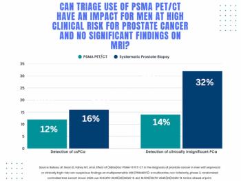
Study: PSMA PET/CT Reduces Biopsy Rate by Nearly 50 Percent for Men with Equivocal or Non-Suspicious Prostate mpMRI
4
FDA Clears AI-Powered Software for Improving Low-Contrast CT Detection
5