- Diagnostic Imaging Vol 31 No 2
- Volume 31
- Issue 2
Game changes with new portable and wireless digital radiography
X-ray, the past and current workhorse in radiology,still garners the majority of all imagingstudies.
X-ray, the past and current workhorse in radiology, still garners the majority of all imaging studies. But when it comes to new advances, the modality tends not to generate the excitement associated with the higher tech alternatives of CT, MR, and PET.
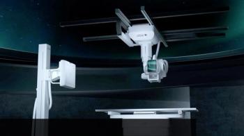
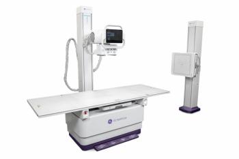
That may have changed a bit with the 2008 RSNA meeting. A new generation of portable digital radiography detectors shouldered its way onto the stage and drew a lot of attention on the exhibit floor. These detectors, which can operate independently both in and outside of the radiography suite, promise new economies for those who have embraced DR and those yet to do so.
They also offer big improvements in workflow and, in many settings, a big improvement in the safety of operations.
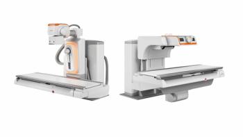
To date, most DR systems have been confined to specific locations and setups, usually a wall- or table-mounted bucky that captures data and sends them to a PACS. Computed radiography provided the flexibility and portability of film while allowing a transition to digital. But unless you had a CR reader where the scan was captured, the techs most often wound up running it back to a place where it could be viewed and then transmitted to the PACS for reading. Without immediate feedback, problems can occur:
• Quality assurance. What if the scan was over- or underexposed, or missed the region of interest? The tech then faced the difficult question of whether to go back and repeat it on a crowded schedule or go ahead and send it through, hoping the radiologist wouldn't complain. If the scan was taken in the intensive care unit, the tech may have forgotten to move aside the ECG leads, thus rendering the image unreadable. Even if CR plates can be read near where they were taken, reading those plates takes time that adds up.
• Patient identification. A variety of systems, usually paper-based and sometimes including yellow sticky notes, were designed to associate the cassette with the patient, but they were not failure proof. Sticky notes or other papers got mixed up as batches of cassettes were processed.
Only recently have DR plates been rugged or light enough to move around. Now they are, and by matching CR's portability, they may offer a good option for many facilities. They can more easily be placed under a bedridden patient's back or held in place to image the head or limb.
Particularly attractive is the notion that they can now go where they are needed, capture images that can be checked for quality in a matter of seconds, and then be transmitted into a PACS.
This digital convenience should also make it possible to more effectively capture patient demographic data and associate it with the image in the PACS. Dr. David Hirschorn, director of informatics at Staten Island University and an editorial advisor for Diagnostic Imaging, said he can envision a scenario where the portable DR system is actually connected to the patient's wrist tag and narrows down the DICOM modality work list to only exams ordered for that patient, preventing the exam from being mislabeled.
This process is not without some challenges. There will be networking issues involved in transmitting data-intensive x-ray images over wireless-or even wired-networks in hospitals. Many hospitals have invested in CR and have thoroughly mastered all the quality and workflow issues involved, so a new investment may not make sense for them. Further, much of what I've described above could be accomplished with portable x-ray machines that have portable wireless CR readers.
Still, it seems fairly clear that the idea of portable and wireless DR detectors marks a significant potential gain and some new thinking about how to manage x-ray imaging in a busy hospital environment. That's an important advance at a time when efficiency and a reduction in medical errors are being demanded.
Articles in this issue
about 17 years ago
International experts, cutting-edgetechnology impress RSNA newbieabout 17 years ago
Radiology gets sent off to the washhouseabout 17 years ago
Clinicians identify tacticsthat minimize risk of NSFabout 17 years ago
Edgy NSF article may have gone too farabout 17 years ago
Study confirms: Digitalmammo lengthens read timeabout 17 years ago
Vertebroplasty data hint atnegative effect on survivalabout 17 years ago
Vampire bat thrombolytic study findings disappointabout 17 years ago
Incidental finding on MRIpoints to multiple sclerosisabout 17 years ago
3D neuro imaging techniquerealizes Orwellian visionabout 17 years ago
Imaging utilization increaseswhere radiologists waneNewsletter
Stay at the forefront of radiology with the Diagnostic Imaging newsletter, delivering the latest news, clinical insights, and imaging advancements for today’s radiologists.


