
|Articles|May 9, 2001
GE recruits Genometrix to combine imaging and in vitro diagnostics
GE Medical Systems has allied with a developer of applied genomics technology, Texas-based Genometrix, to combine genetic and protein-based data with techniques for molecular imaging.The multimodality vendor wants to explore the use of in vitro
Advertisement
GE Medical Systems has allied with a developer of applied genomics technology, Texas-based Genometrix, to combine genetic and protein-based data with techniques for molecular imaging.
The multimodality vendor wants to explore the use of in vitro diagnostics to screen patients for preclinical signs of disease, then follow up with conventional and molecular imaging to monitor at-risk patients, tailor treatment for those with clinical signs of disease, and assess treatment. These tests might involve MRI alone or in combination with other modalities, including CT and nuclear medicine.
The partnership, unveiled April 23 on the exhibit floor of the annual meeting of the International Society for Magnetic Resonance in Medicine in Scotland, will build upon preliminary work already completed by the two companies. Testing, done so far mostly on animals, is expected to expand soon to clinical studies at the University of Texas M.D. Anderson Cancer Center in Houston.
“The vision is to develop methods for detecting life-threatening diseases before symptoms appear and to use those same methods to judge the effect of therapeutic drugs-to see them hitting their targets and determine whether they are effective in a matter of hours rather than weeks,” said Dr. Amit Kakar, manager of MR advanced development at GE. “Being able to see if the patient is resistive to treatment will be a huge point.”
GE and Genometrix are emphasizing the development of methods for identifying precancerous disease. Genomic microarray technologies developed by Genometrix, combined with GE’s high-resolution conventional and molecular modalities using targeted MR contrast agents and MR spectroscopy, show promise, Kakar said. The goal initially will be to detect and quantify risk levels, particularly for breast and prostate cancer. MR spectroscopy will quantitate chemical markers of these cancers. Fine-needle aspiration performed under MRI guidance will extract tissue for definitive genetic testing, which ultimately could provide the basis for tailoring treatment to the patient’s genetic makeup.
“This combination of imaging and genetic testing will change the way we look at disease, diagnose disease, and treat it,” Kakar said.
At first, these techniques may be used to examine patients already treated for cancer in one breast to monitor possible occurrence in the other. MRI might also be used to examine the whole patient in much the same way nuclear medicine whole-body scans are used to identify metastases.
The complementary technologies also have applications outside cancer, Kakar said. GE is developing an MRI technique that promises to distinguish between Alzheimer’s disease and other forms of dementia.
Advertisement
Related Content
Advertisement


Advertisement
Advertisement
Trending on Diagnostic Imaging
1
SNMMI: What Early Research Reveals About the Alpha-Emitting Radioconjugate ATNM-400 for Prostate Cancer
2
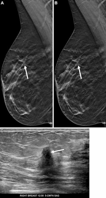
Mammography Study Shows Impact of AI-Powered Slab Reconstruction with DBT
3
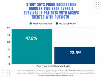
SNMMI: Does Vaccination Enhance Pluvicto Efficacy in mCRPC?
4
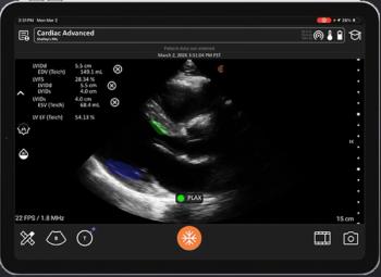
GE HealthCare Unveils 4D Dynamic PET Imaging Software at SNMMI Conference
5